Review
. 2011 Sep 1;25(17):1770-82.
doi: 10.1101/gad.17268411.

Ending the message: poly(A) signals then and now

Affiliations
Review

Ending the message: poly(A) signals then and now

Nick J Proudfoot. Genes Dev. .

Abstract

Polyadenylation [poly(A)] signals (PAS) are a defining feature of eukaryotic protein-coding genes. The central sequence motif AAUAAA was identified in the mid-1970s and subsequently shown to require flanking, auxiliary elements for both 3'-end cleavage and polyadenylation of premessenger RNA (pre-mRNA) as well as to promote downstream transcriptional termination. More recent genomic analysis has established the generality of the PAS for eukaryotic mRNA. Evidence for the mechanism of mRNA 3'-end formation is outlined, as is the way this RNA processing reaction communicates with RNA polymerase II to terminate transcription. The widespread phenomenon of alternative poly(A) site usage and how this interrelates with pre-mRNA splicing is then reviewed. This shows that gene expression can be drastically affected by how the message is ended. A central theme of this review is that while genomic analysis provides generality for the importance of PAS selection, detailed mechanistic understanding still requires the direct analysis of specific genes by genetic and biochemical approaches.


Figures
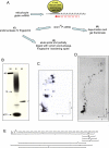
Figure 1.
Figure 1.
(A) Schematic of cDNA synthesis by E. coli DNA polymerase (Klenow subfragment that lacks the 5′–3′exonuclease domain) using oligo(dT)12 as primer (in red), base-paired to mRNA 3′ poly(A) in a reaction mix containing MnCl2 in place of MgCl2 and four deoxyribonucleotides, one α32P-labeled. Short cDNAs produced by this process correspond to the 3′ end of the mRNA 3′ UTR. Purified cDNA was depurinated by formic acid treatment (selectively degrades G and A) releasing oligopyrimidies, especially oligo(dT) copied from the poly(A) sequence of the mRNA. Alternatively cDNA was digested with partially C-specific endonuclease IV, and the endonuclease IV digestion products were then purified and further degraded by partial digestion with venom 5′–3′ exonuclease (Proudfoot 1976). (B) cDNA depurination products were fractionated by denaturing polyacrylamide gel electrophoresis. A series of oligo(dT) products are visible, ranging from dT13–dT50, and correspond to oligo(dT)-primed cDNA generated by oligo(dT), which is base-paired at positions ranging from the 5′ to 3′ ends of the poly(A) tail (known to be about A50 for globin mRNA). Marker (M) and sample (S) lanes are shown, with the positions of oligo(dT)12 and unincorporated α32P dNTP also indicated. (C) 2D fractionation of cDNA endonuclease IV products. The first dimension was by electrophoresis on cellulose acetate strips at pH 3.5. Base composition of oligonucleotide determines mobility. A cellulose acetate strip with fractionated oligonucleotides was blotted onto the bottom of a DEAE cellulose acetate, thin-layer plate using high-salt buffer. Homochromatography was then performed (second dimension) using a buffer (called a homomix) containing partially degraded crude yeast RNA that acts to displace endonuclease IV oligonucleotides from the base of the thin-layer plate. The extent of this displacement depends on molecular size (Brownlee and Sanger 1969). Separated 32P oligonucleotides were then eluted from the thin-layer plate. The first and second dimensions are indicated by arrows. (D) 2D fractionation (as in C) of partial venom exonuclease digested spot 9. A series of products are evident, with adjacent ones varying by 1 nt. The angle of the 2D shift between adjacent spots is characteristic of a specific nucleotide loss. Purine nucleotides (G and A) give a larger shift in the second dimension. T and G give large and small rightward shifts (respectively), while C and A both give small leftward shifts in the first dimension. The sequence TTATT was deduced from observed mobility shifts and corresponds to the PAS AAUAAA. The first T of the PAS cDNA was inferred from depurination data (not shown). (B) Bromophenol blue marker. (E) Separated oligonucleotide sequences were aligned, as some sequences overlap due to partial endonuclease IV digestion. This gave the sequence of the β-globin cDNA adjacent to the poly(A) tail. Endonuclease IV spots not in the β-globin cDNA sequence were inferred to derive from the α-globin cDNA (Proudfoot 1976). († and ‡) Related oligonucleotides from varying 5′ and 3′ ends of cDNA.
Figure 2.
Figure 2.
(A) Sequence alignment of the original six mRNA 3′ ends derived from sequencing technology as outlined in Figure 1. The positions of the conserved AAUAAA signals (boxed) and 3′-terminal nucleotides (underlined) originally noted to be conserved are indicated. (Proudfoot and Brownlee 1976). (B) Current general consensus sequences for the mammalian poly(A) signal. Distance variation between different parts of the PAS is indicated. (Red thunderbolt) Cleavage position.
Figure 3.
Figure 3.
Diagram comparing the mechanism of cotranscriptional 3′-end formation of mammalian polyadenylated mRNA versus unpolyadenylated histone mRNA (RNA is in red, and DNA is in black). For poly(A)+ mRNA, only factors conserved with histone pre-mRNA processing are shown. The rest of the poly(A) complex is depicted by a large grey shadow. Pol II (in light blue) is depicted with striped CTD, and the position of the ssDNA bubble is depicted with associated nascent RNA. Histone 3′ processing factors are indicated. The cap at the 5′ end of mRNA is depicted as a red ball. The histone mRNA 3′ hairpin is shown, as is the U7 RNA hairpin and the interaction with the histone downstream element (HDE). Positions of cis RNA 3′-end processing sequence elements are shown in white boxes. mRNA products of poly(A)+ and histone pre-mRNA 3′-end processing are shown (in boxes), as is the subsequent coupled termination of Pol II by Xrn2-mediated torpedo effects [established for poly(A)+ genes, but only inferred for histone genes].
Figure 4.
Figure 4.
Diagram summarizing the selective use of PAS along a Pol II transcribed gene. PAS in introns are recognized by the poly(A) complex, but this is modulated or blocked by U1snRNP binding to adjacent 5′SS. Recognition of PAS at the end of genes is enhanced by splicing factors bound to the terminal intron 3′SS. Different 3′-terminal PAS are competitively and mutually exclusively used, resulting in APA.

Similar articles

Cited by

References

    1. Adesnik M, Salditt M, Thomas W, Darnell JE 1972. Evidence that all messenger RNA molecules (except histone messenger RNA) contain poly (A) sequences and that the poly(A) has a nuclear function. J Mol Biol 71: 21–30 - PubMed
    1. Amara SG, Jonas V, Rosenfeld MG, Ong ES, Evans RM 1982. Alternative RNA processing in calcitonin gene expression generates mRNAs encoding different polypeptide products. Nature 298: 240–244 - PubMed
    1. Ashe MP, Griffin P, James W, Proudfoot NJ 1995. Poly(A) site selection in the HIV-1 provirus: inhibition of promoter-proximal polyadenylation by the downstream major splice donor site. Genes Dev 9: 3008–3025 - PubMed
    1. Ashe MP, Pearson LH, Proudfoot NJ 1997. The HIV-1 5′ LTR poly(A) site is inactivated by U1 snRNP interaction with the downstream major splice donor site. EMBO J 16: 5752–5763 - PMC - PubMed
    1. Ashfield R, Enriquez-Harris P, Proudfoot NJ 1991. Transcriptional termination between the closely linked human complement genes C2 and factor B: common termination factor for C2 and c-myc? EMBO J 10: 4197–4207 - PMC - PubMed

Publication types

LinkOut - more resources