. 2011 Sep 1;25(17):1835-46.
doi: 10.1101/gad.17175111.

Structural basis for the role of the Sir3 AAA+ domain in silencing: interaction with Sir4 and unmethylated histone H3K79

Affiliations

Structural basis for the role of the Sir3 AAA+ domain in silencing: interaction with Sir4 and unmethylated histone H3K79

Stefan Ehrentraut et al. Genes Dev. .

Abstract

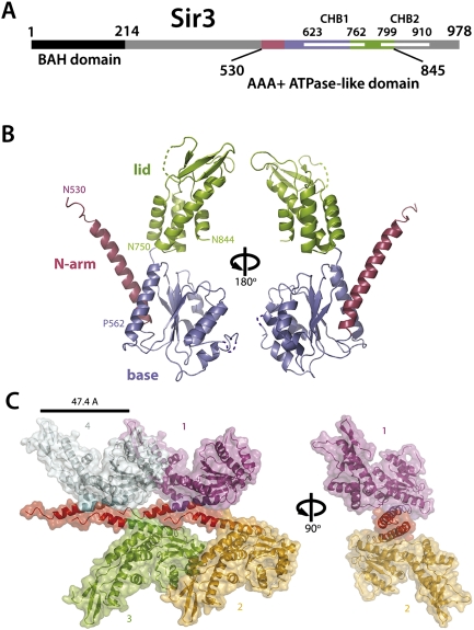
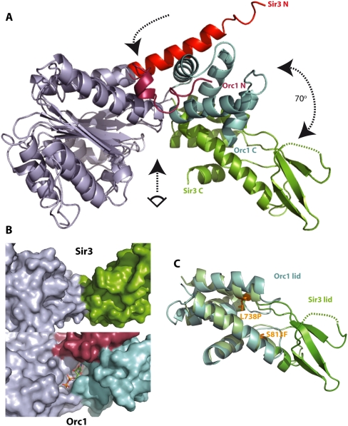
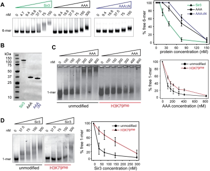
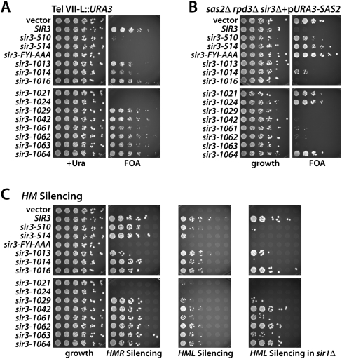
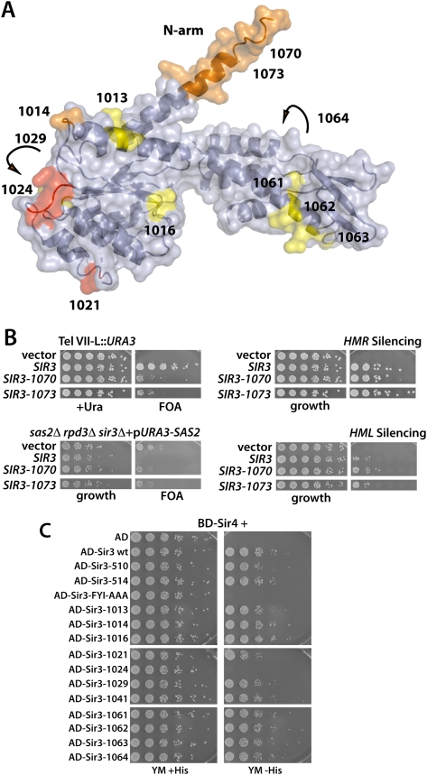
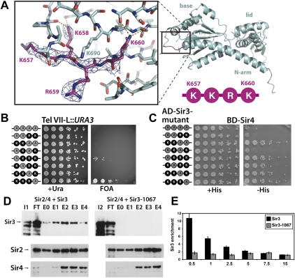
The silent information regulator 2/3/4 (Sir2/3/4) complex is required for gene silencing at the silent mating-type loci and at telomeres in Saccharomyces cerevisiae. Sir3 is closely related to the origin recognition complex 1 subunit and consists of an N-terminal bromo-adjacent homology (BAH) domain and a C-terminal AAA(+) ATPase-like domain. Here, through a combination of structure biology and exhaustive mutagenesis, we identified unusual, silencing-specific features of the AAA(+) domain of Sir3. Structural analysis of the putative nucleotide-binding pocket in this domain reveals a shallow groove that would preclude nucleotide binding. Mutation of this site has little effect on Sir3 function in vivo. In contrast, several surface regions are shown to be necessary for the Sir3 silencing function. Interestingly, the Sir3 AAA(+) domain is shown here to bind chromatin in vitro in a manner sensitive to histone H3K79 methylation. Moreover, an exposed loop on the surface of this Sir3 domain is found to interact with Sir4. In summary, the unique folding of this conserved Sir3 AAA(+) domain generates novel surface regions that mediate Sir3-Sir4 and Sir3-nucleosome interactions, both being required for the proper assembly of heterochromatin in living cells.


Figures

Figure 1.
Figure 1.
The Sir3 AAA+ ATPase-like domain is a structurally divergent family member. (A) Schematic representation of the Sir3 domain structure. (B) Ribbon representation of the Sir3 AAA+ crystal structure spanning amino acids 530–844. The structure is shown in two orientations, rotated through 180°, and colored as in the schematic. Disordered or absent residues are indicated by dashed lines. The relevant subdomains are depicted in red (N-arm), blue (base), and green (lid). Residues marking subdomain boundaries are also indicated. (C) Crystal lattice contacts involving the N-arm helices of four crystallographically related molecules (differently colored, labeled 1–4). The lattice arrangement is depicted in two orientations. Molecules 3 and 4 are omitted for clarity in the right orientation.
Figure 2.
Figure 2.
The Sir3 AAA+ ATPase-like domain has a strikingly different conformation from its closest structural relative, Orc1/cdc6, and does not contain a nucleotide-binding pocket. (A) Superposition of the AAA+-like domains of Sir3 and of S. solfataricus ORC1/cdc6 (2qby, A). Structural alignments were produced by superposition of the residues within the base subdomain of the two structures (RMSD of Cα atoms < 1.6 Å). For clarity, base subdomains are both colored gray. Other subdomain features are colored differently to highlight differences. The arrows indicate the hinge region (top arrow), the rotation between the lid domains of Sir3 and ORC1/Cdc6 (right arrow), and the view into the (potential) nucleotide-binding pocket shown in B (bottom arrow). (B) Surface representation of the presumed nucleotide-binding pocket of Sir3 (top) compared with the ADP bound nucleotide-binding pocket in S. solfataricus ORC1 (bottom). The shallow and wide groove in Sir3 is not compatible with a suggested OAADPR-binding function. (C) Superposition of the lid subdomains of Sir3 and of S. solfataricus ORC1 (2qby, A). Structural alignments were produced by superposition of the helical residues within the lid subdomains of the two structures (RMSD of Cα atoms = 2.1 Å). Additional structural features in the Sir3 lid subdomain are evident (green). These include a threefold anti-parallel β sheet containing mostly positively charged residues. The location of two known point mutants causing a sir3 phenotype (Stone et al. 2000; Buchberger et al. 2008) are also indicated.
Figure 3.
Figure 3.
Binding of the Sir3 AAA+ ATPase-like domain to chromatin was sensitive to methylation of H3K79. (A) The Sir3 protein, the Sir3 AAA+ ATPase-like domain (AAA; amino acids 530–845), or an N-terminal truncation (AAAΔN; amino acids 545–845) was titrated over a constant amount (25 nM) of unmodified 6-mer nucleosomes. (B) SDS-PAGE gel of 1 μg of the Sir3 protein, the Sir3 AAA+ domain, and the N-terminal truncation used in the experiments above (staining with Coomassie brilliant blue). (C) The Sir3 AAA+ ATPase-like domain was titrated over a constant amount (25 nM) of unmodified or H3K79me Cy3-147 mononucleosomes. Samples were separated by native agarose gel electrophoresis, and Cy3-labeled DNA was visualized. The images are representative of at least three independent experiments, and quantifications show the mean value ± SEM of the percent of unbound chromatin compared with the input. (D) Full-length Sir3 was titrated over a constant amount (25 nM) of unmodified or H3K79me mononucleosomes; three independent experiments were analyzed and plotted as described in C.
Figure 4.
Figure 4.
Mutational analysis of the Sir3 AAA+ domain. (A) Telomeric silencing assay with selected sir3 alleles generated in the alanine scan. The SIR3 alleles were introduced into a TEL-VII-L::URA3 sir3Δ strain and tested for their ability to silence the subtelomeric URA3 reporter by plating serial dilutions on 5-FOA plates. Strains were grown for 2 d (5-FOA three) at 30°C. (B) Test of the selected sir3 alleles for their ability to restore the sas2Δ rpd3Δ synthetic lethality. sir3 alleles were introduced into sas2Δ rpd3Δ sir3Δ carrying SAS2 on a URA3-marked plasmid. Strains were serially diluted and tested for their ability to lose the pURA3-SAS2 plasmid on 5-FOA plates. (C) Test of sir3 alleles for their activity in HM silencing. sir3 alleles were introduced into a MATα sir3Δ strain to test for HMR silencing, and into MATa sir3Δ and MATa sir1Δ sir3Δ cells to test for HML silencing. For mating, each dilution of the strains was mixed with 0.3 OD of a mating tester strain of the opposite mating type, spotted on minimal medium for diploid selection, and incubated for 2 d at 30°C. Growth assays for the HML silencing strains showed equal growth of all strains, but were omitted for clarity.
Figure 5.
Figure 5.
Mapping mutant alleles on the Sir3 AAA+ structure. (A) Surface representation of the crystal structure of the Sir3 AAA+-like domain. The positions of the sir3 alleles listed in Table 1 are indicated on the surface in red (strong phenotype), orange (intermediate phenotype), or yellow (weak phenotype). Arrows indicate that the alleles sir3-1029 and sir3-1064 are located on the reverse side of Sir3 in this view. (B) Mutational analysis of the N-terminal arm of Sir3 observed in the crystal structure. Two mutations that partially deleted the N-terminal arm sequence (D amino acids 530–546; sir3-1073) or mutated every amino acid within this region to alanine (sir3-1070) were tested for their effect on telomeric and HM silencing as in Figure 4. (C) The alleles that affected Sir3 function were tested for Sir3–Sir3 and Sir3–Sir4 interaction by a two-hybrid assay. Strains (AEY3055 transformed with the respective plasmids) were tested for activation of the two-hybrid reporter HIS3 by plating serial dilutions on minimal medium with or without histidine.
Figure 6.
Figure 6.
Detailed mutational analysis of the Sir3 residues 657–660. (A) Representation of the Sir3 region spanning the residues K657, K658, R659, and K660. The loop residues 657–660 are shown in stick representation with the respective 2Fo − Fc electron density map contoured at 1.5 σ. Hydrogen bonds formed by the residues are indicated by orange dashed lines. (B) Telomeric silencing assay with the indicated combinations of mutations. Unmodified residues are shown in black, and positions mutated to alanine are shown in gray. The alleles were introduced into a Tel VII-L::URA3 sir3Δ strain and tested for their ability to silence the subtelomeric URA3 reporter as in Figure 4A. (C) Test of the selected sir3 alleles for Sir3–Sir4 two-hybrid interaction as in Figure 5B. (D) In vitro SIR complex assembly with Sir3 and Sir3-1067. Cells infected with Sir2/4, Sir3, or Sir3-1067 expression constructs were lysed, and extracts were mixed. Total extract (I1: extract Sir3; I2: extract Sir3-1067), flow-through (FT), and the eluted fractions were analyzed by Western blotting against HA-tagged Sir3 (top panel), Sir2 (middle panel), and Sir4 (anti-CBP) (bottom panel). (E) Sir3-1067 was unable to bind to telomeres. Sir3 binding at the right telomere of chromosome VI is shown as enrichment in ChIP experiments relative to the enrichment at the control gene SPS2. The amount of enrichment is given as a function of the distance to the telomere end in kilobases. ChIPs were performed with antibodies against HA-Sir3. Error bars give standard deviations.

References

    1. Afonine PV, Mustyakimov M, Grosse-Kunstleve RW, Moriarty NW, Langan P, Adams PD 2010. Joint X-ray and neutron refinement with phenix.refine. Acta Crystallogr D Biol Crystallogr 66: 1153–1163 - PMC - PubMed
    1. Altaf M, Utley RT, Lacoste N, Tan S, Briggs SD, Cote J 2007. Interplay of chromatin modifiers on a short basic patch of histone H4 tail defines the boundary of telomeric heterochromatin. Mol Cell 28: 1002–1014 - PMC - PubMed
    1. Bell SP, Mitchell J, Leber J, Kobayashi R, Stillman B 1995. The multidomain structure of Orc1p reveals similarity to regulators of DNA replication and transcriptional silencing. Cell 83: 563–568 - PubMed
    1. Braunstein M, Rose AB, Holmes SG, Allis CD, Broach JR 1993. Transcriptional silencing in yeast is associated with reduced nucleosome acetylation. Genes Dev 7: 592–604 - PubMed
    1. Buchberger JR, Onishi M, Li G, Seebacher J, Rudner AD, Gygi SP, Moazed D 2008. Sir3–nucleosome interactions in spreading of silent chromatin in Saccharomyces cerevisiae. Mol Cell Biol 28: 6903–6918 - PMC - PubMed

Publication types

MeSH terms

Substances

LinkOut - more resources