. 2011 Sep 1;25(17):1859-70.
doi: 10.1101/gad.15699211.

Human SNM1A and XPF-ERCC1 collaborate to initiate DNA interstrand cross-link repair

Affiliations

Human SNM1A and XPF-ERCC1 collaborate to initiate DNA interstrand cross-link repair

Anderson T Wang et al. Genes Dev. .

Abstract

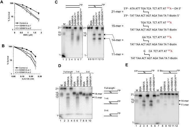
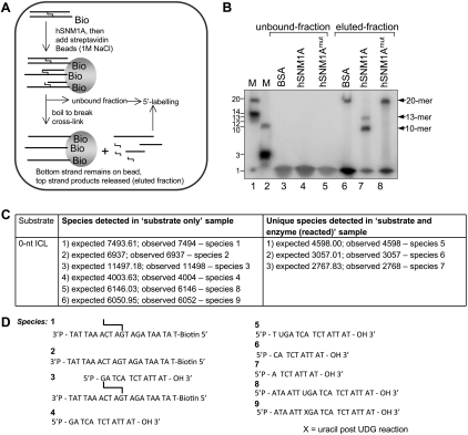
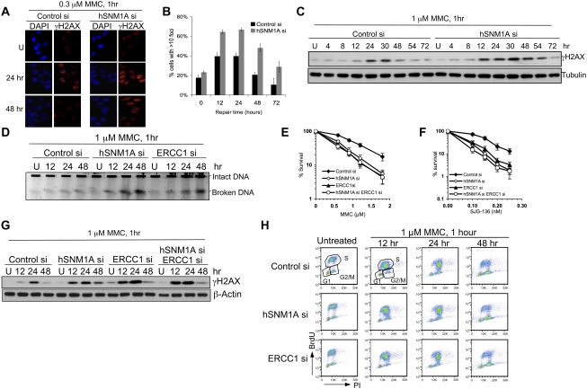
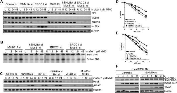
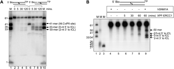
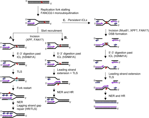
One of the major DNA interstrand cross-link (ICL) repair pathways in mammalian cells is coupled to replication, but the mechanistic roles of the critical factors involved remain largely elusive. Here, we show that purified human SNM1A (hSNM1A), which exhibits a 5'-3' exonuclease activity, can load from a single DNA nick and digest past an ICL on its substrate strand. hSNM1A-depleted cells are ICL-sensitive and accumulate replication-associated DNA double-strand breaks (DSBs), akin to ERCC1-depleted cells. These DSBs are Mus81-induced, indicating that replication fork cleavage by Mus81 results from the failure of the hSNM1A- and XPF-ERCC1-dependent ICL repair pathway. Our results reveal how collaboration between hSNM1A and XPF-ERCC1 is necessary to initiate ICL repair in replicating human cells.


Figures

Figure 1.
Figure 1.
hSNM1A can degrade DNA containing an ICL. (A) A clonogenic survival assay was performed on HeLa cells transfected with two independent siRNAs targeting hSNM1A. Cells were treated with increasing doses of MMC for 1 h. Error bars show standard error of the mean (SEM). (B) Clonogenic survival of siRNA transfected HeLa cells continuously incubated with increasing doses of SJG-136. (C, left panel) Reaction products of hSNM1A on dsDNA (lanes 2–7) or SJG-136 cross-linked dsDNA (lanes 8–13). Hydrolysis was carried out in the absence of protein (lanes 2,8), or with BSA (0.1 μg; lanes 3,9), full-length hSNM1A WT (0.02 μg; lanes 4,10) or its cognate D736A mutant (0.02 μg; lanes 5,11), or E. coli purified ΔN-hSNM1A (hSNM1A-[608–1040]) (0.0005 μg; lanes 6,12) or its cognate D736A mutant (0.0005 μg; lanes 7,13). (Lane 1) 3′-Labeled molecular weight marker oligonucleotides; sizes are indicated. (Right panel) Predicted identity of reaction products obtained based on molecular weight. (D) Reaction (30 min) of hSNM1A with full-length ICL substrate (lanes 2–4), 1-nt “run-up” ICL substrate (lanes 5–7), or 0-nt “run-up” cross-linked dsDNA (lanes 8–10). Hydrolysis was carried out in the presence of BSA (0.1 μg; lanes 2,5,8), full-length hSNM1A (lanes 3,6,9), or its D736A mutant form (lanes 4,7,10). (Lane 1) 3′-Labeled marker oligonucleotides; sizes are indicated. (E) Reaction (30 min) of hSNM1A on cross-linked dsDNA, containing either 5′ phosphate (lanes 2–6) or 5′-biotin (lanes 8–12). Hydrolysis was carried out with BSA (0.1 μg; lanes 2,8), full-length hSNM1A (0.02 μg; lanes 3,9) or its D736A mutant (0.02 μg; lanes 4,10), or ΔN-hSNM1A (0.0005 μg; lanes 5,11) or its D736A mutant (0.0005 μg; lanes 6,12). (Lanes 1,6) 3′-Labeled molecular weight marker oligonucleotides; sizes are indicated.
Figure 2.
Figure 2.
Post-labeling of reaction products at the 5′ end with 32P-γ-ATP suggests that hSNM1A hydrolysis past SJG-136 ICL is exonucleolytic. (A) Schematic diagram of experimental strategy. Substrate bearing a 5′ phosphate was reacted with full-length hSNM1A. The reaction products were immobilized on streptavidin-coated magnetic beads via the biotin moiety on the bottom strand. Boiling the beads releases the top strand product only, which was then post-labeled at the 5′ end with T4 polynucleotide kinase and [32P]-γ-ATP. (B, lanes 1,2) 5′-End-labeled marker oligonucleotides; sizes are as indicated. (Lanes 3–5) Unbound fraction following streptavidin immobilization of biotinylated hSnm1A reaction products labeled with T4PNK and 32P-γ-ATP. (Lanes 6–8) Streptavidin-bound fraction labeled with T4PNK and γ-32P-ATP after boiling to break the cross-link and removal of magnetic beads. The reactions contained either BSA (lanes 3,6) or full-length hSNM1A (lanes 4,7) or its cognate D736A mutant (lanes 5,8). LC-MS confirmed the identity of the hSNM1A reaction products. (C) Experimental and expected masses of the species observed in the experimental mixtures analyzed by mass spectrometry. (D) Structures of the species referred to in C.
Figure 3.
Figure 3.
Depletion of hSNM1A leads to the accumulation of ICL-associated DSBs. (A) HeLa cells transfected with indicated siRNAs were seeded on glass coverslips before treatment with 0.3 μM MMC for 1 h. Cells were fixed at the indicated time points before staining with γH2AX antibodies and DAPI. (B) Quantification of slides from A for percentage of cells with >10 γH2AX foci. Values represent the averages ± SEM of blind scoring from three independent experiments. (C) HeLa cells transfected with indicated siRNAs were treated with 1 μM MMC for 1 h, then incubated in drug-free medium for the indicated time. The levels of γH2AX were determined by immunoblotting. (D) DSB formation was directly detected by PFGE. (E,F) HeLa cells were transfected with control siRNA, siRNAs against hSNM1A, or ERCC1 or in combination. Clonogenic survival assays were carried out as described in Figure 1. (G) γH2AX activation kinetics in cells depleted for hSNM1A, ERCC1, or both factors following MMC treatment. (H) Cell cycle profile after treatment with 1 μM MMC for 1 h. HeLa cells were incubated in drug-free medium after drug treatment and were harvested at the indicated time. Samples were analyzed by FACS, and the cell cycle profiles were presented as BrdU incorporation versus PI staining.
Figure 4.
Figure 4.
Mus81 depletion suppresses γH2AX activation in response to ICLs in HeLa cells. (A) Cells transfected with the indicated siRNAs were treated with 1 μM MMC for 1 h and then incubated in drug-free medium after drug treatment and harvested at the indicated time. Expression levels of proteins were detected by immunoblotting. (B) Amount of broken DNA from cells described in A was analyzed by PFGE. (C) Immunoblotting showing γH2AX levels in cells transfected with the indicated siRNAs and treated with MMC. (D,E) Clonogenic survival of cells transfected with indicated siRNAs following 1 h of MMC treatment. (F) γH2AX, Chk1-ser317 phosphorylation, and FANCD2 monoubiquitination (which produces FANCD2-L) were monitored in control and hSNM1A-depleted cells after MMC.
Figure 5.
Figure 5.
hSNM1A can digest ICL-containing duplex DNA starting from XPF-induced incision. (A) Time course of hSNM1A hydrolysis of blocked (5′-biotinylated) and singly nicked cross-linked substrates by full-length hSNM1A. (Lane 1) 3′-End-labeled marker oligonucleotides; lengths are as indicated. (Lanes 2–5) Hydrolysis of blocked dsDNA 61-mer after 0 min (lane 2), 5 min (lane 3), 30 min (lane 4), or 120 min (lane 5). (Lanes 6–9) Hydrolysis of blocked dsDNA 61-mer treated with Nt.CviPII to introduce a site-specific nick 20 nt from the 5′ end (at nucleotide 41) after 0 min (lane 6), 5 min (lane 7), 30 min (lane 8), or 120 min (lane 9). The guanine that is involved on the ICL on the labeled strand is at position 33 from the 5′-end. (B) Reaction of XPF–ERCC1 and ΔN-hSNM1A on blocked (5′-biotinylated) cross-linked substrate. (Lanes 1–3) 3′-Labeled (lanes 1,3) or 5′-labeled (lane 2) marker oligonucleotides; sizes are indicated. (Lane 5) Unreacted substrate. (Lane 4) Reaction containing ΔN-hSNM1A only. (Lanes 6–8) Reaction containing XPF–ERCC1, stopped after 5 min (lane 6), 30 min (lane 7), or 60 min (lane 8). (Lane 9) Reaction with XPF–ERCC1 for 60 min, followed by reaction with ΔN-hSNM1A for 30 min.
Figure 6.
Figure 6.
Models for context-dependent ICL repair during replication. Following replication fork stalling in the vicinity of the ICL, FANCD2–FANCI becomes activated and in turn orchestrates the repair of ICLs. The repair is initiated by XPF–ERCC1-dependent incisions, either 5′ to the lesion alone or by flanking incisions. Slx4 may help to target XPF–ERCC1 to the site of the ICLs at such structures. Following ICL incision, hSNM1A is able to digest the cross-linked oligonucleotide, leaving a single nucleotide tethered to the complementary strand. The initial incisions could be on either the leading or lagging strand. (A) Should the incision and processing reaction be targeted to the leading strand template, TLS gap filling would produce an intact template for the progression of the nascent leading strand, with a gap remaining in the nascent lagging strand that can be repaired/filled post-replicatively by TLS or HR. (B) Conversely, incision on the lagging strand would allow the leading strand to directly extend past the ICL lesion. Break-induced repair by HR can then take place to re-establish the replication fork. (C) In the situation when two replication forks converge on an ICL, most likely during late S phase, initial incisions could still be produced by XPF–ERCC1, with post-incision processing by hSNM1A. Where XPF–ERCC1 is impaired, the converging fork pathway might predominate due to the failure to initiate repair following the arrival of the first fork, but under such circumstances, we suggest that here Mus81 could be recruited as an alternative means of incising the ICL, generating a two-ended DSB. Following extension of leading strand synthesis past the remaining ICL adduct, HR can restore the replication fork through DSB repair, allowing completion of DNA synthesis.

Similar articles

Cited by

References

    1. Ahkter S, Richie CT, Zhang N, Behringer RR, Zhu C, Legerski RJ 2005. Snm1-deficient mice exhibit accelerated tumorigenesis and susceptibility to infection. Mol Cell Biol 25: 10071–10078 - PMC - PubMed
    1. Akkari YM, Bateman RL, Reifsteck CA, Olson SB, Grompe M 2000. DNA replication is required To elicit cellular responses to psoralen-induced DNA interstrand cross-links. Mol Cell Biol 20: 8283–8289 - PMC - PubMed
    1. Andersson BS, Sadeghi T, Siciliano MJ, Legerski R, Murray D 1996. Nucleotide excision repair genes as determinants of cellular sensitivity to cyclophosphamide analogs. Cancer Chemother Pharmacol 38: 406–416 - PubMed
    1. Aravind L, Walker DR, Koonin EV 1999. Conserved domains in DNA repair proteins and evolution of repair systems. Nucleic Acids Res 27: 1223–1242 - PMC - PubMed
    1. Bae JB, Mukhopadhyay SS, Liu L, Zhang N, Tan J, Akhter S, Liu X, Shen X, Li L, Legerski RJ 2008. Snm1B/Apollo mediates replication fork collapse and S Phase checkpoint activation in response to DNA interstrand cross-links. Oncogene 27: 5045–5056 - PMC - PubMed

Publication types

LinkOut - more resources