. 2011 Sep 20;108(38):15751-6.
doi: 10.1073/pnas.1111498108. Epub 2011 Sep 6.

Function of leukemogenic mixed lineage leukemia 1 (MLL) fusion proteins through distinct partner protein complexes

Affiliations

Function of leukemogenic mixed lineage leukemia 1 (MLL) fusion proteins through distinct partner protein complexes

Debabrata Biswas et al. Proc Natl Acad Sci U S A. .

Abstract

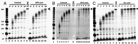
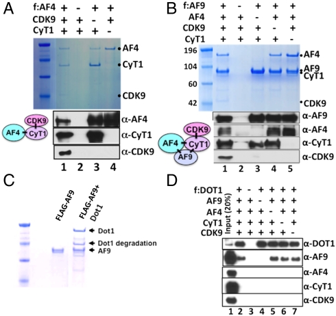
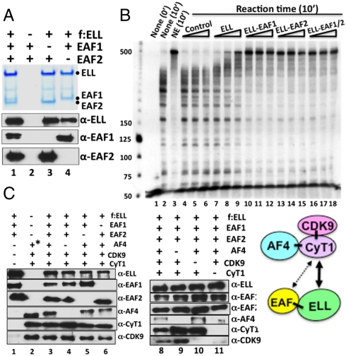
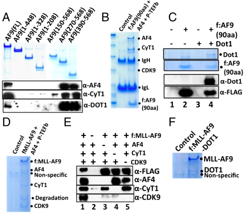
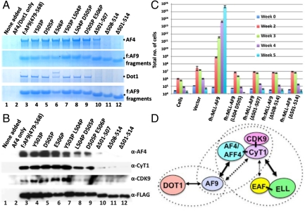
A number of acute leukemias arise from fusion of the mixed lineage leukemia 1 protein (MLL) N terminus to a variety of fusion partners that have been reported to reside in one or more poorly defined complexes linked to transcription elongation through interactions with the histone H3-K79 methyltransferase DOT1 and positive transcription elongation factor b (P-TEFb). Here we first identify natural complexes (purified through fusion partners AF9, AF4, and ELL) with overlapping components, different elongation activities, and different cofactor associations that suggest dynamic interactions. Then, through reconstitution of defined, functionally active minimal complexes, we identify stable subcomplexes that, through newly defined protein-protein interactions, form distinct higher order complexes. These definitive analyses show, for example, that (i) through direct interactions with AF9 and cyclinT1, family members AF4 and AFF4 independently mediate association of P-TEFb with AF9, (ii) P-TEFb, through direct interactions, provides the link for association of ELL and ELL-associated factors 1 and 2 (EAF1 and EAF2) with AF4, and (iii) in the absence of other factors, DOT1 forms a stable complex with AF9 and does not interact with AF9•AF4•P-TEFb complexes. Finally, we show the importance of defined higher order complex formation in MLL-AF9-mediated transcriptional up-regulation and cell immortalization potential in vivo. Thus, our study provides direct mechanistic insight into the role of fusion partners in MLL fusion-mediated leukemogenesis.


Conflict of interest statement

The authors declare no conflict of interest.

Figures

Fig. 1.
Fig. 1.
Functional elongation analyses of AF9-, AF4-, and ELL-associated protein complexes. (A, B, and C) In vitro transcription elongation kinetic assay on a DNA template using the purified AF9 (A), AF4 (B), and ELL (C) complexes (Fig. S1 A, B, and C, respectively). The initial pulse-labeled RNA transcripts (0′ time point) were chased for the indicated time periods to produce runoff transcripts (arrows).
Fig. 2.
Fig. 2.
Baculovirus reconstitution of higher-order protein complexes involving AF4, P-TEFb, AF9, and DOT1. (A) SDS/PAGE and Coomassie blue staining (Upper) and immunoblot (Lower) analyses of the reconstituted AF4•P-TEFb complex and corresponding subunit interactions. Sf9 cells were coinfected, as indicated, with baculoviruses that express FLAG-AF4 (f:AF4), His-cyclinT1 (CyT1), and His-Cdk9 and complexes were purified on M2 agarose. (B) SDS/PAGE and Coomassie blue staining (Upper) and immunoblot (Lower) analyses of the reconstituted AF9•AF4•P-TEFb complex and corresponding subunit interactions. (C) SDS/PAGE and Coomassie blue staining of the reconstituted AF9•DOT1 complex. (D) Immunoblot analysis indicating that DOT1 and the AF4•P-TEFb complex do not interact directly or assemble into a common complex containing AF9.
Fig. 3.
Fig. 3.
Baculovirus reconstitution of a functional ELL•EAF1/2 complex and its interaction with the AF4•P-TEFb complex. (A) SDS/PAGE, Coomassie blue staining (Upper) and immunoblot (Lower) analyses of the reconstituted ELL•EAF1/2 complex and corresponding subunit interactions. (B) In vitro transcription elongation assay (as described in Fig. 1) with purified baculovirus-expressed ELL and with purified reconstituted ELL•EAF1/2 complexes (Fig. 3A). (C) Immunoblot analysis of the reconstituted ELL•EAF1/2•AF4•P-TEFb complex and corresponding subunit interactions. The asterisk indicates protein complex purification through FLAG-AF4 in this lane.
Fig. 4.
Fig. 4.
Mapping of an AF9 domain that interacts directly with other factors: Implication for MLL–AF9-mediated higher order complex assembly. (A) SDS/PAGE and Coomassie blue staining showing expression of FLAG-AF9 fragments (Upper) and immunoblot showing interactions of these fragments with coexpressed AF4, cyclinT1, and DOT1 (Lower). (B and C) SDS/PAGE and Coomassie blue staining of complexes reconstituted with AF9 (90-amino acid fragment from 479–568) and either AF4•P-TEFb (B) or DOT1 (C). (D) SDS/PAGE and Coomassie blue staining of the reconstituted MLL–AF9•AF4•P-TEFb complex. (E) Immunoblot analysis of the corresponding subunit interactions in the MLL–AF9•AF4•P-TEFb complex. (F) SDS/PAGE Coomassie blue staining of the reconstituted MLL–AF9•DOT1 complex.
Fig. 5.
Fig. 5.
Mutational analysis of AF9: A role for higher order complex assembly in MLL–AF9-mediated transformation. (A) Analysis of direct AF4 and DOT1 interactions with the small AF9 fragment (90 amino acids) and derived mutants. In two independent experiments, Sf9 cells were infected with baculoviruses expressing FLAG-AF9 fragments and either AF4 (Top and Second Rows) or DOT1 (Third and Bottom Rows) and corresponding complexes were purified on M2 agarose and analyzed by SDS/PAGE with Coomassie blue staining. (B) Analysis of higher order complex assembly between the small AF9 fragment or mutant derivatives and the AF4•P-TEFb complex. (C) Hematopoietic transformation assays with retroviruses carrying MLL–AF9 or the indicated mutants. (D) Diagram showing possible functionally active higher order complex formations involving AF9, AF4, AFF4, and ELL along with interacting partners DOT1, P-TEFb, EAF1, and EAF2. The dashed ovals represent the stable complexes observed in the reconstitution studies with defined factors. Note that the AF9•DOT1 and AF9•AF4•P-TEFb complexes are mutually exclusive.

References

    1. Krivtsov AV, Armstrong SA. MLL translocations, histone modifications and leukaemia stem-cell development. Nat Rev Cancer. 2007;7:823–833. - PubMed
    1. Marschalek R. Mechanisms of leukemogenesis by MLL fusion proteins. Br J Haematol. 2011;152:141–154. - PubMed
    1. Huret JL, Dessen P, Bernheim A. An atlas of chromosomes in hematological malignancies. Example: 11q23 and MLL partners. Leukemia. 2001;15:987–989. - PubMed
    1. Krivtsov AV, et al. H3K79 methylation profiles define murine and human MLL–AF4 leukemias. Cancer Cell. 2008;14:355–368. - PMC - PubMed
    1. Milne TA, Martin ME, Brock HW, Slany RK, Hess JL. Leukemogenic MLL fusion proteins bind across a broad region of the Hox a9 locus, promoting transcription and multiple histone modifications. Cancer Res. 2005;65:11367–11374. - PubMed

Publication types

MeSH terms

LinkOut - more resources