. 2011 Sep 20;108(38):15864-9.
doi: 10.1073/pnas.1112672108. Epub 2011 Sep 6.

Neural-specific elongation of 3' UTRs during Drosophila development

Affiliations

Neural-specific elongation of 3' UTRs during Drosophila development

Valérie Hilgers et al. Proc Natl Acad Sci U S A. .

Abstract

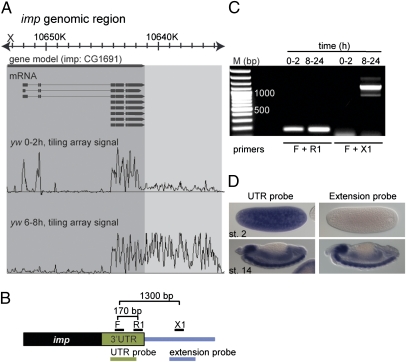
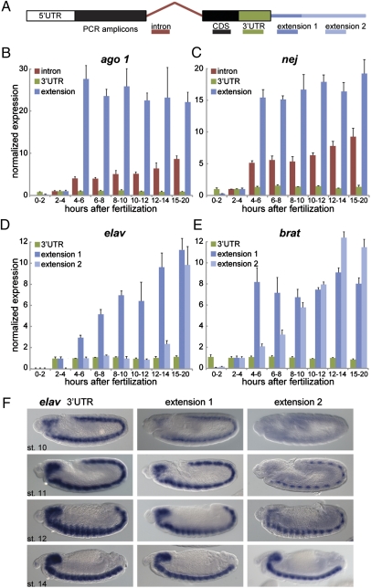
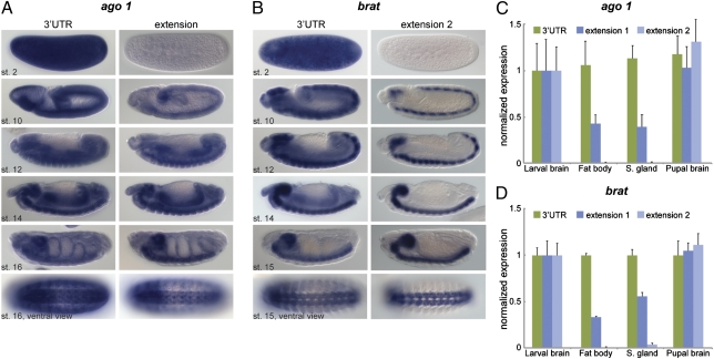
The 3' termini of eukaryotic mRNAs influence transcript stability, translation efficiency, and subcellular localization. Here we report that a subset of developmental regulatory genes, enriched in critical RNA-processing factors, exhibits synchronous lengthening of their 3' UTRs during embryogenesis. The resulting UTRs are up to 20-fold longer than those found on typical Drosophila mRNAs. The large mRNAs emerge shortly after the onset of zygotic transcription, with several of these genes acquiring additional, phased UTR extensions later in embryogenesis. We show that these extended 3' UTR sequences are selectively expressed in neural tissues and contain putative recognition motifs for the translational repressor, Pumilio, which also exhibits the 3' lengthening phenomenon documented in this study. These findings suggest a previously unknown mode of posttranscriptional regulation that may contribute to the complexity of neurogenesis or neural function.


Conflict of interest statement

The authors declare no conflict of interest.

Figures

Fig. 1.
Fig. 1.
Extended 3′ UTRs. (A) Representative, whole-genome tiling array data for total Drosophila RNA from age-staged embryos produced by Manak et al. (17). Displayed is the signal from 0–2 h AF and 6–8 h AF embryos for the gene imp, aligned to the gene model. The extent of the annotated transcripts and the extent of the observed 3′ UTR extension are highlighted in dark and light gray, respectively. (B) Schematic representation of the 3′ end of imp mRNA. Indicated are the positions of PCR primers designed to amplify a fragment of the annotated imp 3′ UTR (170 bp) or a fragment consisting of 400 bp of 3′ UTR and 900 bp of the presumed extension (1,300 bp). At the bottom, probes used in D were designed to hybridize to the 3′ UTR or to the presumed extension (not to scale). (C) RT-PCR on total RNA from early (0–2 h AF) and late (8-24 h AF) embryos using the primers represented in B. The imp 3′ UTR extension can only be detected later in embryogenesis, whereas the annotated imp transcript is detected at both time points. (D) In situ hybridization for imp using probes represented in B. The 3′ UTR extension is detected only in later embryos, whereas the annotated transcript is both maternally deposited and zygotically expressed. In all figures, anterior is to the left and dorsal is at the top.
Fig. 2.
Fig. 2.
Timing of 3′ UTR isoform expression during embryogenesis. (A) Schematic of qRT-PCR experiments. Primers were designed to amplify target sequences in the intron (red), coding region (CDS; black), 3′ UTR (green), and proximal (extension 1; dark blue) or distal (extension 2; light blue) portions of the extension. Note that the primer pairs “intron,” “CDS,” and “UTR” detect both the short and the long forms, whereas “extension” primer pairs are specific to the long form. (B–E) Quantification of indicated transcripts by qRT-PCR using primer combinations shown in A. RNA was extracted from embryos at different times in embryonic development (indicated in h AF). At indicated time points, expression levels were calculated for individual primer sets by normalization to rp49 mRNA (constitutively expressed). Baseline expression was established by setting the value at 2–4 h AF to 1. Represented is the normalized expression relative to levels obtained with the CDS primer pair. The increase in extension levels compared with CDS levels indicates an increase in the ratio of the long form to the short form at later time points, whereas UTR sequences remain unchanged compared with the coding region. Error bars represent mean ± SD of three to four biological replicates for each time point. (F) In situ hybridization for elav using probes against the 3′ UTR (Left) or an extension (Center and Right) at indicated embryonic stages (st.).
Fig. 3.
Fig. 3.
Neural enrichment of long 3′ UTR isoforms. (A and B) In situ hybridization for ago1 (A) and brat (B) using probes that hybridize to the coding region and 3′ UTR (Left) or an extension (Right). (C and D) Quantification of ago1 (C) and brat (D) transcripts by qRT-PCR using primer combinations shown in Fig. 2A. RNA was extracted from brains, fat body, and salivary glands of third-instar larvae as well as from pupal brains (48 h after puparium formation). Levels were normalized to rp49 mRNA, and expression in larval brains was set to the value 1 for each primer pair. Represented is the fold change relative to levels obtained with the CDS primer pair. Error bars represent mean ± SD of four to six biological replicates for each tissue.

References

    1. Sonoda J, Wharton RP. Drosophila Brain Tumor is a translational repressor. Genes Dev. 2001;15:762–773. - PMC - PubMed
    1. Murata Y, Wharton RP. Binding of Pumilio to maternal hunchback mRNA is required for posterior patterning in Drosophila embryos. Cell. 1995;80:747–756. - PubMed
    1. Irish V, Lehmann R, Akam M. The Drosophila posterior-group gene nanos functions by repressing hunchback activity. Nature. 1989;338:646–648. - PubMed
    1. Wharton RP, Struhl G. RNA regulatory elements mediate control of Drosophila body pattern by the posterior morphogen nanos. Cell. 1991;67:955–967. - PubMed
    1. Wreden C, Verrotti AC, Schisa JA, Lieberfarb ME, Strickland S. Nanos and pumilio establish embryonic polarity in Drosophila by promoting posterior deadenylation of hunchback mRNA. Development. 1997;124:3015–3023. - PubMed

Publication types

MeSH terms

LinkOut - more resources