. 2011 Sep 20;108(38):15828-33.
doi: 10.1073/pnas.1111312108. Epub 2011 Sep 6.

Targeted killing of a mammalian cell based upon its specialized metabolic state

Affiliations

Targeted killing of a mammalian cell based upon its specialized metabolic state

Peter B Alexander et al. Proc Natl Acad Sci U S A. .

Abstract

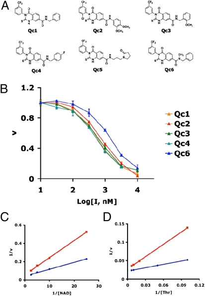
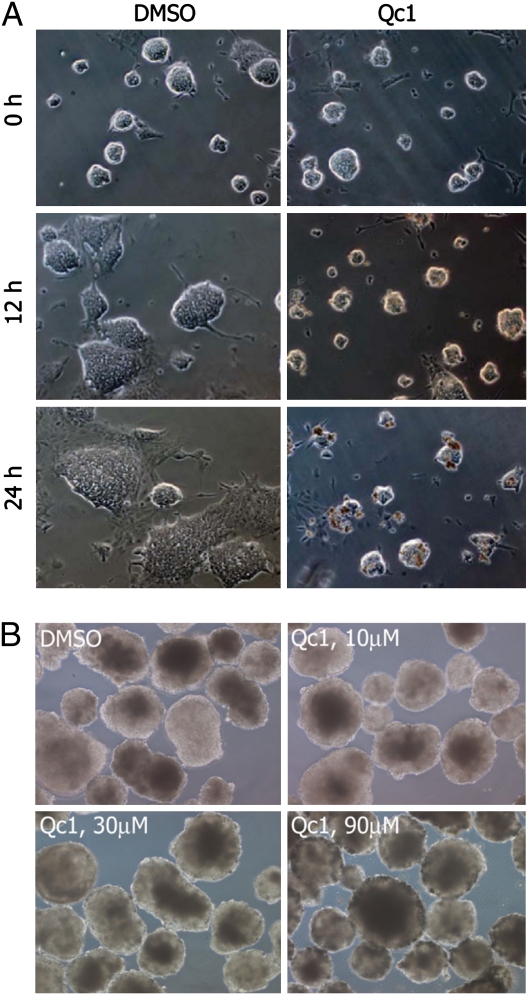
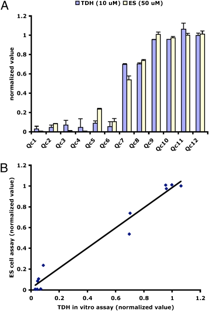
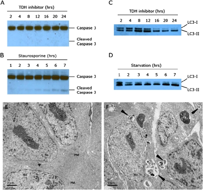
Mouse ES cells use a mitochondrial threonine dehydrogenase (TDH) enzyme to catabolize threonine into glycine and acetyl-CoA. Measurements of mRNA abundance have given evidence that ES cells express upwards of 1,000-fold higher levels of TDH mRNA than any of seven other mouse tissues tested. When cell culture medium is deprived of threonine, ES cells rapidly discontinue DNA synthesis, arrest cell division, and eventually die. Such studies led to the conclusion that mouse ES cells exist in a threonine-dependent metabolic state. Proceeding with the assumption that the active TDH enzyme should be essential for the growth and viability of mouse ES cells, we performed a drug screen in search of specific inhibitors of the purified TDH enzyme. Such efforts led to the discovery of a class of quinazolinecarboxamide (Qc) compounds that inhibit the ability of the TDH enzyme to catabolize threonine into glycine and acetyl-CoA. Administration of Qc inhibitors of TDH to mouse ES cells impeded cell growth and resulted in the induction of autophagy. By contrast, the same chemicals failed to affect the growth of HeLa cells at concentrations 300-fold higher than that required to kill mouse ES cells. It was likewise observed that the Qc class of TDH inhibitors failed to affect the growth or viability of ES cell-derived embryoid body cells known to have extinguished TDH expression. These studies demonstrate how it is possible to kill a specific mammalian cell type on the basis of its specialized metabolic state.


Conflict of interest statement

The authors declare no conflict of interest.

Figures

Fig. 1.
Fig. 1.
Chemical structures and potency of TDH inhibitors. (A) Structures of the six best hits from the TDH inhibitor screen. The compounds contained a Qc scaffold with various peripheral modifications. (B) IC50 values were determined by titrating the compounds from 10 nM to 10 μM and measuring TDH activity (Methods). The approximate IC50 for all six compounds was 0.5 μM. (C and D) Lineweaver-Burk analysis of enzyme inhibition. TDH activity was assayed in the absence and presence of the Qc inhibitor at the NAD+ and threonine concentrations shown. Blue curves depict data obtained in the absence of inhibitor, and red curves depict data obtained in the presence of inhibitor. Both Vmax (y intercept) and Km (x intercept) were altered in the presence of inhibitor, indicative of mixed noncompetitive inhibition.
Fig. 2.
Fig. 2.
Effect of TDH inhibition on ES cell growth, colony morphology, and embryoid body morphology. (A) Feederless ES cells (E14 strain) were cultured on glass chamber slides and imaged using phase contrast microscopy. When treated with vehicle (DMSO) alone, ES cell colonies rapidly grew in size. Upon exposure to the TDH inhibitor, ES cells failed to proliferate, with colony size remaining unchanged for the first 12 h. After 24 h, clusters of densely packed cells became apparent at the surface of the colonies, indicative of cell death. (B) E14 ES cells were grown in suspension without Leukemia Inhibitory Factor (LIF) for 10 d to allow differentiation into embryoid bodies. Embryoid bodies were then treated for 24 h with vehicle or Qc1 at indicated concentrations.
Fig. 3.
Fig. 3.
Correlation between TDH inhibition and ES cell cytotoxicity for 12 Qc compounds. (A) Twelve structurally related Qc compounds were assayed for their ability to inhibit TDH in vitro (10-μM compound) and impair proliferation of ES cells (50-μM compound). The normalized values for both assays are plotted in histogram form. Qc1–Qc6 are the TDH inhibitors identified through the small molecule screen, and Qc7–Qc12 are structurally related compounds found in the UTSWMC chemical library that were not identified in the screen. Qc1–Qc6 are potent TDH inhibitors that kill ES cells with EC50 ≈3 μM. Qc7 and Qc8 are weak TDH inhibitors with EC50 >50 μM. Qc9–Qc12 did not inhibit TDH in vitro and displayed no toxicity when added to the ES cell growth medium. (B) Same as in A, with data plotted as an xy scatter plot.
Fig. 4.
Fig. 4.
Accumulation of threonine and AICAR and depletion of acetyl-CoA and mTHF in ES cells treated with TDH inhibitors. Feederless ES cells were treated with 10 μM of the Qc1 TDH inhibitor for 0, 1, 2, 3, and 4 h before extraction of metabolites in 50% aqueous methanol and subsequent LC-MS/MS analysis. Metabolites increasing in abundance as a function of exposure to the Qc1 inhibitor of TDH are shown in red. Metabolites decreasing in abundance are shown in green.
Fig. 5.
Fig. 5.
TDH inhibition results in elevated autophagy but not apoptosis. (A) Protein lysates from ES cells treated with 10 μM TDH inhibitor were extracted in 0.5% Nonidet P-40, separated using SDS/PAGE, and immuoblotted using an antibody specific for caspase 3. Despite visible cell death, no cleaved caspase 3 could be detected after 24 h of TDH inhibitor treatment. (B) Control Western blot showing cleaved caspase 3 in response to treatment with 1 μM staurosporine. (C) ES cell lysates were immunoblotted with an antibody to LC3. After 16 h of TDH inhibitor treatment, most of the LC3 protein was in the LC3-II (lipidated) form, indicative of increased autophagic activity. (D) Control Western blot showing increased autophagy in response to nutrient starvation. ES cells were cultured in HBSS and harvested at the times indicated. (E and F) Electron microscopy of ES cells treated with TDH inhibitor. Mouse ES cells were cultured on plastic coverslips for 24 h with or without TDH inhibitor. Cells were fixed with 2.5% glutaraldehyde in 0.1 M cacodylate buffer and embedded in Embed-812 Resin. Thin sections (70–90 nm in thickness) were stained with 2% aqueous uranyl acetate and lead citrate and examined by transmission electron microscopy. (E) Control ES cells treated with DMSO only. (F) ES cells cultured in the presence of 10 μM TDH inhibitor. Arrowheads indicate autophagic compartments. Nu, nucleus; PM, plasma membrane; Ly, dense lysosomes.

References

    1. Smyth HF, Carpenter CP. Water-soluble derivatives of p-aminobenzene-sulfonamide (sulfanilamide) Science. 1938;87:350–351. - PubMed
    1. Roland S, Ferone R, Harvey RJ, Styles VL, Morrison RW. The characteristics and significance of sulfonamides as substrates for Escherichia coli dihydropteroate synthase. J Biol Chem. 1979;254:10337–10345. - PubMed
    1. Tally FP, Sutter VL, Finegold SM. Metronidazole versus anaerobes. In vitro data and initial clinical observations. Calif Med. 1972;117:22–26. - PMC - PubMed
    1. Carson LE, Campbell CC. The inhibitory effect of three antihistamine compounds on the growth of fungi pathogenic for man. Science. 1950;111:689–691. - PubMed
    1. Nyfeler R, Keller-Schierlein W. [Metabolites of microorganisms. 143. Echinocandin B, a novel polypeptide-antibiotic from Aspergillus nidulans var. echinulatus: Isolation and structural components] Helv Chim Acta. 1974;57:2459–2477. - PubMed

Publication types

MeSH terms