. 2011 Sep 20;108(38):16104-9.
doi: 10.1073/pnas.1107533108. Epub 2011 Sep 6.

Prostaglandin E2 release from astrocytes triggers gonadotropin-releasing hormone (GnRH) neuron firing via EP2 receptor activation

Affiliations

Prostaglandin E2 release from astrocytes triggers gonadotropin-releasing hormone (GnRH) neuron firing via EP2 receptor activation

Jerome Clasadonte et al. Proc Natl Acad Sci U S A. .

Abstract

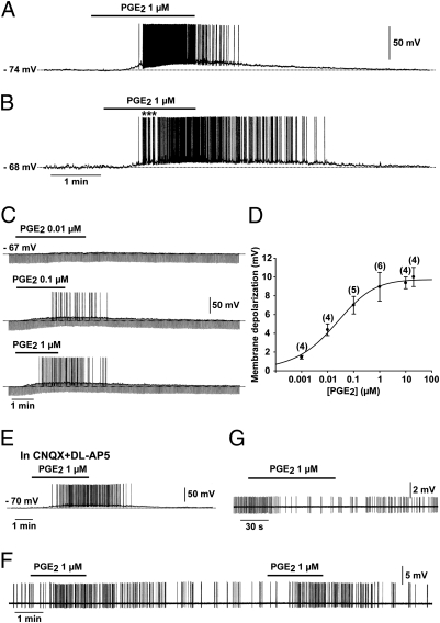
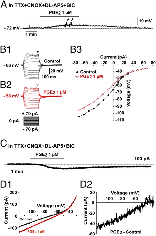
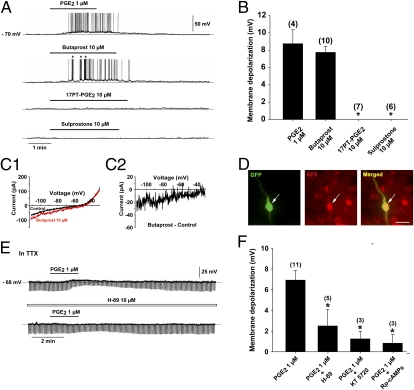
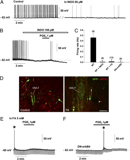
Astrocytes in the hypothalamus release prostaglandin E(2) (PGE(2)) in response to cell-cell signaling initiated by neurons and glial cells. Upon release, PGE(2) stimulates the secretion of gonadotropin-releasing hormone (GnRH), the neuropeptide that controls reproduction, from hypothalamic neuroendocrine neurons. Whether this effect on GnRH secretion is accompanied by changes in the firing behavior of these neurons is unknown. Using patch-clamp recording we demonstrate that PGE(2) exerts a dose-dependent postsynaptic excitatory effect on GnRH neurons. These effects are mimicked by an EP2 receptor agonist and attenuated by protein kinase A (PKA) inhibitors. The acute blockade of prostaglandin synthesis by indomethacin (INDO) or the selective inhibition of astrocyte metabolism by fluoroacetate (FA) suppresses the spontaneous firing activity of GnRH neurons in brain slices. Similarly, GnRH neuronal activity is reduced in mice with impaired astrocytic PGE(2) release due to defective erbB signaling in astrocytes. These results indicate that astrocyte-to-neuron communication in the hypothalamus is essential for the activity of GnRH neurons and suggest that PGE(2) acts as a gliotransmitter within the GnRH neurosecretory system.


Conflict of interest statement

The authors declare no conflict of interest.

Figures

Fig. 1.
Fig. 1.
PGE2 powerfully activates GnRH neurons. (A and B) Whole-cell current-clamp recordings showing the effect of bath application of PGE2 on two GnRH neurons. Note that in these two silent GnRH neurons, which had a resting membrane potential of −74 mV (A) and −68 mV (B), respectively, PGE2 induced a reversible membrane depolarization that led to the initiation of spike firing. The effect was short-lived in A and long-lasting in B. Note that in B, the spike firing started with a bursting pattern (*). (C) Whole-cell current-clamp recording of a single GnRH neuron showing that PGE2 (0.01–1 μM) depolarized the membrane in a dose-dependent manner. Note that in this cell, 0.01 μM of PGE2 did not trigger spike firing. In this and the following figures, the downward deflections correspond to voltage responses to 300 ms hyperpolarizing current pulses used to test the membrane input resistance. (D) Dose–response curve of the PGE2-induced membrane depolarization. The numbers of neurons tested at each dose is given in parentheses. Error bars indicate SEM. The EC50 for the PGE2-induced membrane depolarization was 0.018 μM, based on a logistic equation fitted to the data points. (E) The excitatory effect of PGE2 on GnRH neurons persisted in the presence of the AMPA/kainate and NMDA receptor antagonists CNQX (20 μM) and DL-AP5 (100 μM), respectively. (F) Loose patch-clamp recording showing the excitatory effect of PGE2 on a GnRH neuron, characterized by a reversible acceleration of firing. Note that the effect was reproduced by a second application of PGE2 to the same neuron. (G) Loose patch-clamp recording showing the inhibitory effect of PGE2 on a non-GnRH neuron located in the vicinity of GnRH-GFP neurons. The effect was characterized by a reversible slowing down of firing.
Fig. 2.
Fig. 2.
The PGE2-induced activation of GnRH neurons is direct and involves an inward current. (A) Whole-cell current-clamp recording showing that PGE2 depolarized GnRH neurons in the presence of TTX (0.5 μM), CNQX (20 μM), DL-AP5 (100 μM), and bicuculline (BIC, 20 μM). Arrowheads indicate the time of application of hyperpolarizing and depolarizing current pulses to trace current–voltage relationships. (B1B3) Responses of a GnRH neuron to current injection from −70 pA to +70 pA before (B1, control) and during application of PGE2 (B2). The current–voltage relationship (B3) for the corresponding neuron obtained before (control) and during the application of PGE2 indicate that PGE2 decreased the slope of the linear part of the curve, indicating a decrease in the membrane input resistance. Note that the two curves converged at −40 mV in this example. (C) Whole-cell voltage-clamp recording showing that PGE2 evoked an inward current in GnRH neurons in the presence of TTX (0.5 μM), CNQX (20 μM), DL-AP5 (100 μM), and BIC (20 μM). The inward current was recorded at a holding potential of −70 mV. (D1 and D2) Current traces evoked by voltage ramps (duration, 12.5 s) from −120 mV to −30 mV at a holding potential of −70 mV in GnRH neurons. (D1) Traces showing the current responses to the voltage ramp in GnRH neurons before (control) and during application of PGE2. (D2) PGE2-induced current obtained after subtracting the control from the PGE2 curve. Note that the PGE2-induced current is suppressed at −40 mV.
Fig. 3.
Fig. 3.
The PGE2-induced activation of GnRH neurons is mediated by the EP2 receptor and requires the cAMP/PKA pathway. (A) In a single GnRH neuron recorded with a whole-cell current clamp, the EP2 receptor agonist, butaprost, evoked a membrane depolarization similar to that induced by PGE2, whereas the EP1 receptor agonist 17-phenyl trinor PGE2 (17PT-PGE2) and the EP1–3 receptor agonist, sulprostone, had no effect. Note that the discharge elicited by the butaprost-induced membrane depolarization led to bursts of action potentials (*). (B) Bar graph illustrating the membrane depolarization in GnRH neurons induced by PGE2, butaprost, 17PT-PGE2, and sulprostone (*P < 0.05 compared with the membrane depolarization induced by PGE2, one-way ANOVA; n = 4–10 neurons). Error bars indicate SEM. (C1 and C2) Current traces evoked by voltage ramps (duration, 12.5 s) from −120 mV to −30 mV at a holding potential of −70 mV in GnRH neurons. (C1) Traces showing the current response to the voltage ramp in GnRH neurons before (control) and during application of butaprost, a selective EP2 receptor agonist. (C2) Butaprost-induced current obtained after subtracting the control from the butraprost curve. Note that the butraprost-induced current was suppressed at −35 mV in this example. (D) EP2 receptor immunoreactivity (red) was detected in the cell body of GnRH-GFP neurons (green, arrow). (Scale bar, 20 μm.) (E) Whole-cell current-clamp recording of a single GnRH neuron in the presence of TTX (0.5 μM) showing the effect of PGE2 in the absence (Upper) and presence (Lower) of the membrane-permeable PKA inhibitor H89. Note that H89 attenuated the membrane depolarization and the decrease in membrane input resistance (downward deflections) induced by PGE2. (F) Bar graph illustrating the membrane depolarization induced by PGE2 alone or in the presence of H89 and two other membrane-permeable compounds, the more selective PKA inhibitor KT 5720 and the cAMP antagonist Rp-cAMPs (*P < 0.05 compared with the membrane depolarization induced by PGE2 alone, one-way ANOVA; n = 3–11 neurons). Error bars indicate SEM.
Fig. 4.
Fig. 4.
Astrocytic prostaglandin production sustains the electrical activity of GnRH neurons. Recordings of GnRH neurons were performed under whole-cell current-clamp using pipette solution 2 (ps2) (Materials and Methods) to obtain a background of spontaneous activity from GnRH neurons. (A) GnRH neurons showed spontaneous activity (control), which was reduced after 10 min of perfusion with indomethacin (INDO; 50 μM), an inhibitor of the cyclooxygenases, enzymes responsible for prostaglandin production. (B) INDO at a higher concentration (100 μM) strongly attenuated the spontaneous activity of GnRH neurons, accompanied by membrane hyperpolarization. Note that PGE2 reversed the inhibitory effect of INDO. (C) Bar graph illustrating the firing rate of GnRH neurons recorded in brain slices from wild-type mice (WT) exposed to INDO or fluoroacetate (FA) and from DN-erbB4 mice. (*P < 0.05 compared with the firing rate of GnRH neurons recorded in brain slices from WT mice, one-way ANOVA; n = 6–12 neurons). Error bars indicate SEM. (D) Pretreatment of brain slices with fluoroacetate (FA, 5 mM, 60–120 min), a glial toxin, impaired the astrocytic uptake of sulforhodamine101 (SR101, red). Arrows show cells that took up SR101 in the vicinity of GnRH neurons (GFP, green) under control conditions (Left). After FA treatment, very few cells were labeled with SR101 (Right, arrowhead). Images were acquired at the level of the organum vasculosum of the lamina terminalis (OVLT). (Scale bar, 100 μm.) (E) FA strongly reduced the firing rate of GnRH neurons. In this example, the slice was pretreated with FA for 60 min and action potentials could be driven by a brief injection of a depolarizing current (*). Note that PGE2 retained its depolarizing effect. (F) In DN-erbB4 mice, in which astrocytic PGE2 release is diminished, most GnRH neurons were silent. In this recording, action potentials could be driven by a brief injection of depolarizing current (*). Note that PGE2 retained its depolarizing effect and reduced the membrane input resistance (downward deflections).

Similar articles

Cited by

References

    1. Gordon GR, et al. Astrocyte-mediated distributed plasticity at hypothalamic glutamate synapses. Neuron. 2009;64:391–403. - PMC - PubMed
    1. Henneberger C, Papouin T, Oliet SH, Rusakov DA. Long-term potentiation depends on release of D-serine from astrocytes. Nature. 2010;463:232–236. - PMC - PubMed
    1. Panatier A, et al. Glia-derived D-serine controls NMDA receptor activity and synaptic memory. Cell. 2006;125:775–784. - PubMed
    1. Gourine AV, et al. Astrocytes control breathing through pH-dependent release of ATP. Science. 2010;329:571–575. - PMC - PubMed
    1. Halassa MM, et al. Astrocytic modulation of sleep homeostasis and cognitive consequences of sleep loss. Neuron. 2009;61:213–219. - PMC - PubMed

Publication types

MeSH terms