. 2011 Sep 20;108(38):15954-9.
doi: 10.1073/pnas.1108460108. Epub 2011 Sep 6.

Reactibodies generated by kinetic selection couple chemical reactivity with favorable protein dynamics

Affiliations

Reactibodies generated by kinetic selection couple chemical reactivity with favorable protein dynamics

Ivan Smirnov et al. Proc Natl Acad Sci U S A. .

Abstract

Igs offer a versatile template for combinatorial and rational design approaches to the de novo creation of catalytically active proteins. We have used a covalent capture selection strategy to identify biocatalysts from within a human semisynthetic antibody variable fragment library that uses a nucleophilic mechanism. Specific phosphonylation at a single tyrosine within the variable light-chain framework was confirmed in a recombinant IgG construct. High-resolution crystallographic structures of unmodified and phosphonylated Fabs display a 15-Å-deep two-chamber cavity at the interface of variable light (V(L)) and variable heavy (V(H)) fragments having a nucleophilic tyrosine at the base of the site. The depth and structure of the pocket are atypical of antibodies in general but can be compared qualitatively with the catalytic site of cholinesterases. A structurally disordered heavy chain complementary determining region 3 loop, constituting a wall of the cleft, is stabilized after covalent modification by hydrogen bonding to the phosphonate tropinol moiety. These features and presteady state kinetics analysis indicate that an induced fit mechanism operates in this reaction. Mutations of residues located in this stabilized loop do not interfere with direct contacts to the organophosphate ligand but can interrogate second shell interactions, because the H3 loop has a conformation adjusted for binding. Kinetic and thermodynamic parameters along with computational docking support the active site model, including plasticity and simple catalytic components. Although relatively uncomplicated, this catalytic machinery displays both stereo- and chemical selectivity. The organophosphate pesticide paraoxon is hydrolyzed by covalent catalysis with rate-limiting dephosphorylation. This reactibody is, therefore, a kinetically selected protein template that has enzyme-like catalytic attributes.


Conflict of interest statement

The authors declare no conflict of interest.

Figures

Fig. 1.
Fig. 1.
Chemical structures of compounds used in the study: p-nitrophenyl 8-methyl-8-azabicyclo[3.2.1]octyl phenylphosphonate (1b; R = H) and its biotinylated derivative (1a; R = biotinyl); diisopropyl fluorophosphate (DFP; 2); 4-(2-aminoethyl)benzenesulfonyl fluoride (AEBSF; 3); 2-diethoxyphosphorylthioethyl-trimethylammonium iodide (echothiophate; 4); O,O-diethyl O-(4-nitrophenyl) phosphate (paraoxon; 5); O-(4-nitrophenylphosphoryl) choline (6).
Fig. 2.
Fig. 2.
Structural analysis of reactibody FabA.17. Crystallographic snapshots of the two-chamber active center. α-Chain trace renderings on the upper (A) and lower (B) chambers and merged (C) of the substrate binding site of the A.17 active site shown in space-filling mode. The light chain is shown in pale blue, and the heavy chain is shown in green for A–C. A top view showing aromatic residues forming the walls of the upper cavity and lower chamber centered on reactive side chain of Y-L37 in the unmodified (D) and phosphonate-modified (E) structures. The light chain is shown in gray, the heavy chain is in blue, the VH loop is in magenta, and the OP molecule is in green. Key residues are represented as sticks, with nitrogen atoms marked by deep blue, oxygen atoms marked by red, and phosphorus marked by orange. A bound chloride ion, indicated by the green sphere, is constant to modification (Fig. S3A).
Fig. 3.
Fig. 3.
Comparison of active site cavities of natural and de novo created biocatalysts. Chemically selected reactibody FabA.17 has a deep substrate binding niche. Cross-section views of the active center of esterolytic antibodies 49G7, TEPC15, aldolase antibody 33F12, choline esterases AChE and BChE, and antibody A.17 complexed with their ligands. In each case, the distance measured is the height of a pyramid with a triangle base constructed on the three residues nearest to the entrance of the active site, and apexes are the residue nearest to the ligand.
Fig. 4.
Fig. 4.
Superposition of active sites of native (green) and OP-modified (blue) FabA.17 (A), illustrating the conformational changes of Y-L37, W- L92, F-L100, and residues of the VH loop (H-H104, N-H105, A-H107, and W-H109). Phosphonate moiety is in red. (B) Representative view of the active site of 1b-modified A.17 Fab showing a strong H bond between a nonbridging phosphonyl oxygen and water molecule w614 (2.55 Å). H-Y34 stabilizes a cluster of six water molecules by participating in their H-bond network. The light chain is in gray, the heavy chain is in blue, and the OP molecule is in green. Hydrogen bonds are represented by gray dashes. A bound chloride ion, indicated by the green sphere, is independent of modification (Fig. S4).
Fig. 5.
Fig. 5.
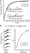
Discrimination of the modification mechanism between A.17 reactibody and phosphonate 1b. Experimental and fitted kinetic curves of interaction of A.17 with 1b. The quality of fit of kinetic models to the experimental data was assessed by monitoring residuals against time for different scheme fits (A). Presteady state kinetics curves of the intrinsic Trp fluorescence changing of A.17 depending on concentration of phosphonate 1b (B Left). Dependence of kobs of the 1b interaction with reactibody A.17 on substrate concentration (B Right). A Inset shows the reaction mechanism used for data fitting and elementary constant calculation.
Fig. 6.
Fig. 6.
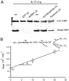
Description of multistep covalent catalysis by A.17 reactibody. Covalent reactivity of A.17 and its mutants with biotinylated phosphonate 1a. Antibodies were incubated for 1 h at 37 °C with PBS buffer alone or 1 mM each of 2 (DFP) and 3 (AEBSF) and for 16 h with PBS buffer alone or 1 mM each 4 (echothiophate) and 5 (paraoxon). All samples were then incubated for 1 h at 37 °C with 100 μM of 1a and analyzed by Western blot (A). Concentrations of antibodies were normalized as confirmed by comparable staining of heavy chains on the same blots (A Upper). Three-step reaction of A.17 antibody with 5 in the presence of NH2OH (B).

References

    1. Benkovic SJ, Hammes-Schiffer S. A perspective on enzyme catalysis. Science. 2003;301:1196–1202. - PubMed
    1. Tawfik DS. Messy biology and the origins of evolutionary innovations. Nat Chem Biol. 2010;6:692–696. - PubMed
    1. Gupta RD, et al. Directed evolution of hydrolases for prevention of G-type nerve agent intoxication. Nat Chem Biol. 2011;7:120–125. - PubMed
    1. Belogurov A, Jr, Kozyr A, Ponomarenko N, Gabibov A. Catalytic antibodies: Balancing between Dr. Jekyll and Mr. Hyde. Bioessays. 2009;31:1161–1171. - PubMed
    1. Lerner RA, Benkovic SJ, Schultz PG. At the crossroads of chemistry and immunology: Catalytic antibodies. Science. 1991;252:659–667. - PubMed

Publication types

MeSH terms

LinkOut - more resources