

Comparative transcriptional study of the putative mannose donor biosynthesis genes in virulent Mycobacterium tuberculosis and attenuated Mycobacterium bovis BCG strains
- PMID: 21896775
- PMCID: PMC3257929
- DOI: 10.1128/IAI.05635-11
Comparative transcriptional study of the putative mannose donor biosynthesis genes in virulent Mycobacterium tuberculosis and attenuated Mycobacterium bovis BCG strains
Abstract
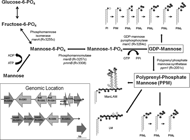
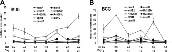
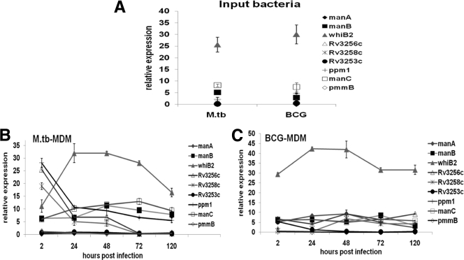
Mycobacterium tuberculosis contains mannosylated cell wall components which are important in macrophage recognition and response. The building block for the mannosyl constituents of these components is GDP-mannose, which is synthesized through a series of enzymes involved in the mannose donor biosynthesis pathway. Nothing is known about the expression levels of the genes encoding these enzymes during the course of infection. To generate transcriptional profiles for the mannose donor biosynthesis genes from virulent M. tuberculosis and attenuated Mycobacterium bovis BCG, bacteria were grown in broth culture and within human macrophages. Our results with broth-grown bacteria show that there are differences in expression of the selected genes between M. tuberculosis and BCG, with increased expression of manC in M. tuberculosis and manA in BCG during stationary-phase growth. Results for M. tuberculosis extracted from within macrophages show that whiB2 is highly expressed and manB and manC are moderately expressed during infection. Rv3256c, Rv3258c, and ppm1 have high expression levels early and decreased expression as the infection progresses. Results with BCG show that, as in M. tuberculosis, whiB2 is highly expressed throughout infection, whereas there is either low expression or little change in expression of the remaining genes studied. Overall, our results show that there is differential regulation of expression of several genes in the mannose donor biosynthesis pathway of M. tuberculosis and BCG grown in broth and within macrophages, raising the possibility that the level of mannose donors may vary during the course of infection and thereby impact the biosynthesis of mannose-containing cell wall molecules.
Figures
Similar articles
-
The Impact of Genome Region of Difference 4 (RD4) on Mycobacterial Virulence and BCG Efficacy.Front Cell Infect Microbiol. 2017 Jun 8;7:239. doi: 10.3389/fcimb.2017.00239. eCollection 2017. Front Cell Infect Microbiol. 2017. PMID: 28642843 Free PMC article.
-
Characterization of the transcriptional regulator Rv3124 of Mycobacterium tuberculosis identifies it as a positive regulator of molybdopterin biosynthesis and defines the functional consequences of a non-synonymous SNP in the Mycobacterium bovis BCG orthologue.Microbiology (Reading). 2010 Jul;156(Pt 7):2112-2123. doi: 10.1099/mic.0.037200-0. Epub 2010 Apr 8. Microbiology (Reading). 2010. PMID: 20378651 Free PMC article.
-
Evidence for occurrence of the ESAT-6 protein in Mycobacterium tuberculosis and virulent Mycobacterium bovis and for its absence in Mycobacterium bovis BCG.Infect Immun. 1996 Jan;64(1):16-22. doi: 10.1128/iai.64.1.16-22.1996. Infect Immun. 1996. PMID: 8557334 Free PMC article.
-
Mycobacterium bovis lipids: virulence and vaccines.Vet Microbiol. 2011 Jul 5;151(1-2):91-8. doi: 10.1016/j.vetmic.2011.02.030. Epub 2011 Feb 24. Vet Microbiol. 2011. PMID: 21420803 Review.
-
Genomics of Mycobacterium bovis.Tuberculosis (Edinb). 2001;81(1-2):157-63. doi: 10.1054/tube.2000.0269. Tuberculosis (Edinb). 2001. PMID: 11463237 Review.
Cited by
-
Lipoglycans contribute to innate immune detection of mycobacteria.PLoS One. 2011;6(12):e28476. doi: 10.1371/journal.pone.0028476. Epub 2011 Dec 2. PLoS One. 2011. PMID: 22164297 Free PMC article.
-
Mycobacterium tuberculosis universal stress protein Rv2623 interacts with the putative ATP binding cassette (ABC) transporter Rv1747 to regulate mycobacterial growth.PLoS Pathog. 2017 Jul 28;13(7):e1006515. doi: 10.1371/journal.ppat.1006515. eCollection 2017 Jul. PLoS Pathog. 2017. PMID: 28753640 Free PMC article.
-
Inflammation and Metabolism of Influenza-Stimulated Peripheral Blood Mononuclear Cells From Adults With Obesity Following Bariatric Surgery.J Infect Dis. 2022 Dec 28;227(1):92-102. doi: 10.1093/infdis/jiac345. J Infect Dis. 2022. PMID: 35975968 Free PMC article.
-
Underestimated Manipulative Roles of Mycobacterium tuberculosis Cell Envelope Glycolipids During Infection.Front Immunol. 2019 Dec 18;10:2909. doi: 10.3389/fimmu.2019.02909. eCollection 2019. Front Immunol. 2019. PMID: 31921168 Free PMC article. Review.
-
Normalization of the levels of inflammatory molecules in Mycobacterium smegmatis-infected U937 cells by fibrate pretreatment.Biol Res. 2014 Sep 15;47(1):42. doi: 10.1186/0717-6287-47-42. Biol Res. 2014. PMID: 25299393 Free PMC article.
References
-
- Alam M. S., Garg S. K., Agrawal P. 2009. Studies on structural and functional divergence among seven WhiB proteins of Mycobacterium tuberculosis H37Rv. FEBS J. 276:76–93 - PubMed
-
- Briken V., Porcelli S. A., Besra G. S., Kremer L. 2004. Mycobacterial lipoarabinomannan and related lipoglycans: from biogenesis to modulation of the immune response. Mol. Microbiol. 53:391–403 - PubMed
-
- Ganguly N., Siddiqui I., Sharma P. 2008. Role of M. tuberculosis RD-1 region encoded secretory proteins in protective response and virulence. Tuberculosis (Edinb.) 88:510–517 - PubMed
-
- Garcia Pelayo M. C., et al. 2009. Gene expression profiling and antigen mining of the tuberculin production strain Mycobacterium bovis AN5. Vet. Microbiol. 133:272–277 - PubMed
Publication types
MeSH terms
Substances
Grants and funding
LinkOut - more resources
Full Text Sources
Molecular Biology Databases
Miscellaneous
