. 2011 Nov;31(21):4286-97.
doi: 10.1128/MCB.05668-11. Epub 2011 Sep 6.

IRE1-dependent activation of AMPK in response to nitric oxide

Affiliations

IRE1-dependent activation of AMPK in response to nitric oxide

Gordon P Meares et al. Mol Cell Biol. 2011 Nov.

Abstract

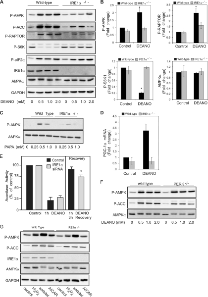
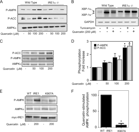
While there can be detrimental consequences of nitric oxide production at pathological concentrations, eukaryotic cells have evolved protective mechanisms to defend themselves against this damage. The unfolded-protein response (UPR), activated by misfolded proteins and oxidative stress, is one adaptive mechanism that is employed to protect cells from stress. Nitric oxide is a potent activator of AMP-activated protein kinase (AMPK), and AMPK participates in the cellular defense against nitric oxide-mediated damage in pancreatic β-cells. In this study, the mechanism of AMPK activation by nitric oxide was explored. The known AMPK kinases LKB1, CaMKK, and TAK1 are not required for the activation of AMPK by nitric oxide. Instead, this activation is dependent on the endoplasmic reticulum (ER) stress-activated protein IRE1. Nitric oxide-induced AMPK phosphorylation and subsequent signaling to AMPK substrates, including Raptor, acetyl coenzyme A carboxylase, and PGC-1α, is attenuated in IRE1α-deficient cells. The endoribonuclease activity of IRE1 appears to be required for AMPK activation in response to nitric oxide. In addition to nitric oxide, stimulation of IRE1 endoribonuclease activity with the flavonol quercetin leads to IRE1-dependent AMPK activation. These findings indicate that the RNase activity of IRE1 participates in AMPK activation and subsequent signaling through multiple AMPK-dependent pathways in response to nitrosative stress.


Figures

Fig. 1.
Fig. 1.
Nitric oxide activates AMPK and modifies mTOR signaling. (A) INS832/13 cells were treated with the nitric oxide donor DEANO for 0 to 60 min (B) or the indicated concentrations of AICAR for 1 h. (C) INS832/13 cells were mock transfected or transfected with siRNA for AMPKα1 and AMPKα2 (together) for 48 h followed by treatment with DEANO (1 mM) for the indicated times. (D) INS832/13 cells were transduced with an adenoviral dominant negative AMPK using a multiplicity of infection (MOI) of 50 to 200 for 24 h followed by treatment with 1 mM DEANO for 30 min. Following treatments, the cells were harvested, and lysates were separated by SDS-gel electrophoresis followed by Western blot analysis using the antibodies specific for the indicated target proteins. Results are representative of three independent experiments.
Fig. 2.
Fig. 2.
LKB1, CaMKK, and TAK1 are not essential for nitric oxide-induced activation of AMPK. (A) INS832/13 cells were mock transfected or transfected with two distinct siRNAs for LKB1 for 48 h followed by treatment with 100 μM H2O2 or 1 mM DEANO for 30 min. (B) INS832/13 cells were pretreated for 30 min with 1 to 10 μM concentrations of the CaMKK inhibitor STO-609 followed by treatment with 1 mM DEANO for 30 min. (C) INS832/13 cells were mock transfected or transfected with siRNA for TAK1 for 48 h followed by treatment with 1 mM DEANO for 30 min. (D) INS832/13 cells were transfected with siRNA for LKB1 and/or TAK1 for 48 h or pretreated for 30 min with 10 μM STO-609 as indicated. Cells were then treated with 1 mM DEANO for 30 min, and target protein levels were analyzed by Western blot analysis. (E) Summary of the effects of target molecule inhibition on DEANO-induced AMPK phosphorylation (*, 30 μM PKCζ attenuated AMPK phosphorylation but was toxic to cells, and the loss of AMPK phosphorylation was not confirmed by siRNA for PKCζ). Results are representative of three independent experiments.
Fig. 3.
Fig. 3.
IRE1 participates in the activation of AMPK by nitric oxide. (A) INS832/13 cells were treated with 1 mM DEANO for the indicated times or left untreated, followed by Western blot analysis for phosphorylated PERK and eIF2α. (B) INS832/13 were mock transfected (−) or transfected with siRNA for IRE1α for 48 h, followed by treatment with the indicated concentrations of DEANO for 30 min. The cells were harvested, and AMPK phosphorylation and total IRE1α and AMPK levels were determined by Western blot analysis. (C) The effects of a 30-min treatment with 1 mM DEANO on XBP1 splicing in INS832/13 cells transfected with IRE1α siRNA are shown. Splicing was analyzed by qRT-PCR. (D) INS832/13 cells, mock transfected or transfected with IRE1α siRNA as for panel B, were treated with the indicated concentrations of AICAR for 30 min. The cells were harvested, and levels of phosphorylated AMPK and ACC and total levels of AMPK and IRE1α were determined by Western blot analysis. Results are representative of two or three individual experiments.
Fig. 4.
Fig. 4.
Nitric oxide-induced AMPK activation and signaling is attenuated in IRE1-deficient cells. (A) Wild-type and IREα−/− MEFs were treated with the indicated concentrations of DEANO for 10 min, and the phosphorylation of AMPK, ACC, Raptor, S6K, and eIF2α was determined by Western blot analysis. Total levels of IRE1α confirm its absence in the deficient MEFs, and total levels of AMPK and GAPDH are shown as loading controls. (B) The phosphorylated forms of AMPK, Raptor, and S6K1 and total AMPK in wild-type and IRE1α-deficient MEFs treated with 1 mM DEANO for 10 min were quantified. (C) Wild-type and IREα−/− cells were treated with the indicated concentrations of PAPA for 15 min, and levels of phosphorylated AMPK and total AMPK were determined by Western blot analysis. (D) Wild type and IREα−/− cells were treated with 1 mM DEANO for 3 h and PGC-1α mRNA accumulation was determined by qRT-PCR. (E) INS832/13 cells were transfected with scrambled (control) or IRE1α siRNA for 24 h and then either treated for 1 h with DEANO or treated for 1 h with DEANO, washed, and cultured for 3 additional hours. The cells were isolated, and mitochondrial aconitase activity was measured. (F) Wild-type or PERK−/− MEFs were treated with the indicated concentrations of DEANO for 10 min, and levels of phosphorylated AMPK and ACC and total AMPK were determined by Western blot analysis. (G) Wild-type and IREα−/− MEFs were treated with the AMPK activator H2O2 (100 μM) or sorbitol (0.6 M) for 30 min or AICAR (2 mM) for 2 h. The MEFs were harvested, and levels of phosphorylated AMPK and ACC and total levels of IRE1α, AMPK, and GAPDH were determined by Western blot analysis. Data are means ± standard errors of the means (SEM) (B, D, and E; n = 3 to 4; *, P < 0.05) or are representative of three independent experiments (A, C, F, and G).
Fig. 5.
Fig. 5.
IRE1α RNase but not ER stress activates AMPK. (A) Wild-type MEFs were treated with tunicamycin (5 μM) or DEANO (1 mM) for the indicated times, and XBP-1 splicing was measured by RT-PCR. (B) Wild-type MEFs were treated with 5 μM tunicamycin for the indicated times or left untreated, and phosphorylated AMPK, Raptor, and eIF2α were examined by Western blot analysis. Wild-type MEFs treated with DEANO were used as a positive control. The asterisk indicates a variable cross-reactive band. (C) INS832/13 cells were treated with thapsigargin (1 μM) or DTT (2 mM) followed by examination of eIF2α phosphorylation; DEANO was used as a positive control. (D and E) INS832/13 cells transfected with wild-type IRE1α or with IRE1α-K599A (kinase deficient) or IRE1α-K907A (RNase deficient) mutants were treated with DEANO (1 mM) for 30 min, and AMPK phosphorylation was examined by Western blot. (F) Stable INS1 cell lines were treated for 16 h with doxycycline (1 μg/ml) to induce WT or K907A-IRE1α-myc, followed by treatment with IL-1β (10 U/ml) for 16 h. The level of phosphorylated AMPK was quantified. Data are means ± SEM; n = 3; *, P < 0.05 versus WT control.
Fig. 6.
Fig. 6.
Quercetin stimulates IRE1α RNase-dependent AMPK activation. (A) Wild-type and IREα−/− MEFs were treated with the indicated concentrations of quercetin, and phosphorylated AMPK and ACC were measured by Western blotting. (B) Wild-type and IREα−/− MEFs were treated with tunicamycin (5 μM) or quercetin (200 μM) for 2 h, and XBP-1 splicing was measured by RT-PCR. XBP-1u and XBP-1s indicate unspliced and spliced XBP-1, respectively. (C and D) INS832/13 cells were treated with the indicated concentrations of quercetin, and phosphorylated AMPK and ACC were measured by Western blotting. (E and F) Stable INS1 cell lines were treated for 16 h with doxycycline (1 μg/ml) to induce WT or K907A-IRE1α-myc, followed by treatment with quercetin for 1 h. The levels of phosphorylated AMPK and total AMPK and IRE1α-myc were examined. Data are means ± SEM (D and F; n = 3; *, P < 0.05 versus control) or are representative of three independent experiments (A, B, C, and E).
Fig. 7.
Fig. 7.
Schematic diagram of nitric oxide-activated AMPK activation in β cells. Nitric oxide induces IRE1-dependent phosphorylation of AMPK and subsequent AMPK-dependent phosphorylation of Raptor and ACC. The AMPK signaling node connects IRE1 to mTORC1 in response to nitric oxide (black arrows) and is independent of the currently known AMPK kinases (gray arrows, indicating that they do not participate in this pathway). The direct effects of nitric oxide on IRE1 are currently unknown.

Similar articles

Cited by

References

    1. Ahn J., Lee H., Kim S., Park J., Ha T. 2008. The anti-obesity effect of quercetin is mediated by the AMPK and MAPK signaling pathways. Biochem. Biophys. Res. Commun. 373:545–549 - PubMed
    1. Almeida A., Moncada S., Bolaños J. 2004. Nitric oxide switches on glycolysis through the AMP protein kinase and 6-phosphofructo-2-kinase pathway. Nat. Cell Biol. 6:45–51 - PubMed
    1. Arnush M., Scarim A., Heitmeier M., Kelly C., Corbett J. 1998. Potential role of resident islet macrophage activation in the initiation of autoimmune diabetes. J. Immunol. 160:2684–2691 - PubMed
    1. Bhaskar P. T., Hay N. 2007. The two TORCs and Akt. Dev. Cell 12:487–502 - PubMed
    1. Calabrese V., et al. 2009. Nitric oxide in cell survival: a janus molecule. Antioxid. Redox. Signal. 11:2717–2739 - PubMed

Publication types

MeSH terms

LinkOut - more resources