

Exploratory study on pathogenesis of far-eastern spotted fever
- PMID: 21896812
- PMCID: PMC3163874
- DOI: 10.4269/ajtmh.2011.10-0660
Exploratory study on pathogenesis of far-eastern spotted fever
Abstract
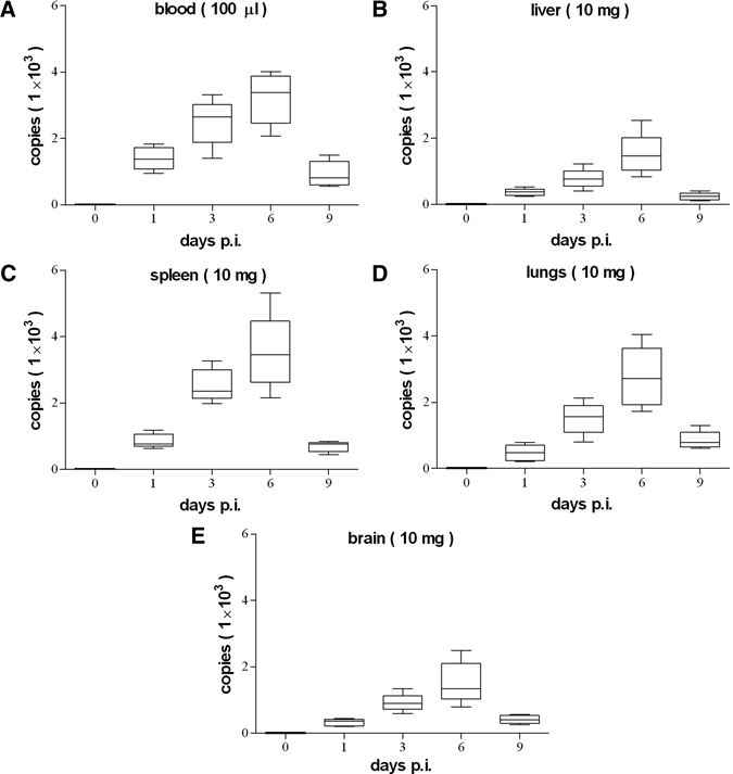
Far-eastern spotted fever is an emerging disease caused by Rickettsia heilongjiangensis, a tick-borne obligate intracellular bacterium. In this study, R. heilongjiangensis was used to infect BALB/c mice by inoculation of retro-orbital venous plexus to imitate a blood infection caused by tick biting. We found that R. heilongjiangensis rapidly entered the circulation for systemic dissemination and the pathogen existed in liver, spleen, lungs, and brain of the mice at least 9 days post-infection (p.i.). Severe pathological lesions were observed in liver, lungs, and brain at Day 6 p.i. In addition, the elevated levels of inflammatory cytokines, including interferon-γ, tumor necrosis factor, and CC chemokine, were detected in the infected organs at Day 3 p.i. Our results reveal that R. heilongjiangensis may cause an infection in BALB/c mice and the pathological lesions in the infected mice are associated with host inflammatory response induced by R. heilongjiangensis.
Figures


References
-
- Raoult D, Berbis P, Roux V, Xu W, Maurin M. A new tick-transmitted disease due to Rickettsia slovaca. Lancet. 1997;350:112–113. - PubMed
-
- Uchida T, Uchiyama T, Kumano K, Walker DH. Rickettsia japonica sp. nov., the etiological agent of spotted fever group rickettsiosis in Japan. Int J Syst Bacteriol. 1992;42:303–305. - PubMed
-
- Wu YM, Zhang ZQ, Wang HJ, Yang Q, Feng L, Wang JW. Investigation on the epidemiology of Far-East tick-borne spotted fever in the northeastern area of China. Zhonghua Liu Xing Bing Xue Za Zhi. 2008;29:1173–1175. - PubMed
Publication types
MeSH terms
Substances
LinkOut - more resources
Full Text Sources
