. 2011 Nov;60(11):2922-7.
doi: 10.2337/db11-0220. Epub 2011 Sep 6.

Deletion of the G6pc2 gene encoding the islet-specific glucose-6-phosphatase catalytic subunit-related protein does not affect the progression or incidence of type 1 diabetes in NOD/ShiLtJ mice

Affiliations

Deletion of the G6pc2 gene encoding the islet-specific glucose-6-phosphatase catalytic subunit-related protein does not affect the progression or incidence of type 1 diabetes in NOD/ShiLtJ mice

James K Oeser et al. Diabetes. 2011 Nov.

Abstract

Objective: Islet-specific glucose-6-phosphatase catalytic subunit-related protein (IGRP), now known as G6PC2, is a major target of autoreactive T cells implicated in the pathogenesis of type 1 diabetes in both mice and humans. This study aimed to determine whether suppression of G6p2 gene expression might therefore prevent or delay disease progression.

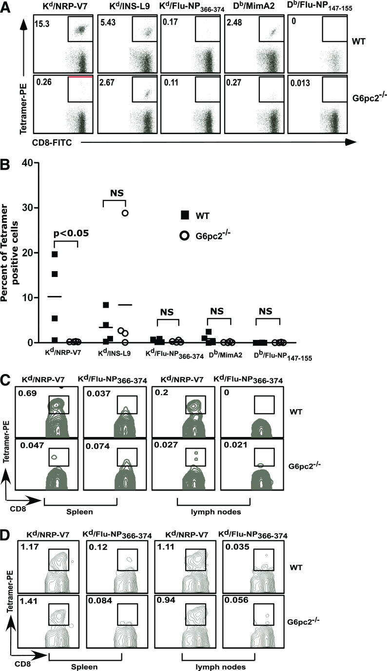
Research design and methods: G6pc2(-/-) mice were generated on the NOD/ShiLtJ genetic background, and glycemia was monitored weekly up to 35 weeks of age to determine the onset and incidence of diabetes. The antigen specificity of CD8(+) T cells infiltrating islets from NOD/ShiLtJ G6pc2(+/+) and G6pc2(-/-) mice at 12 weeks was determined in parallel.

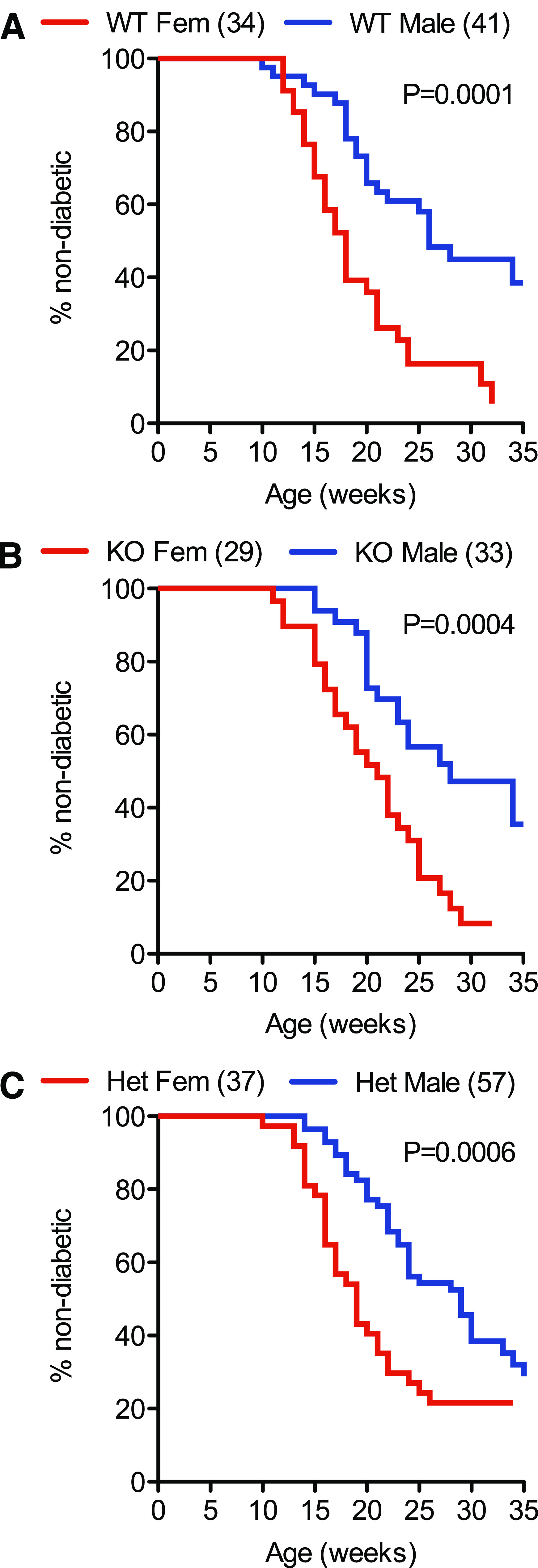
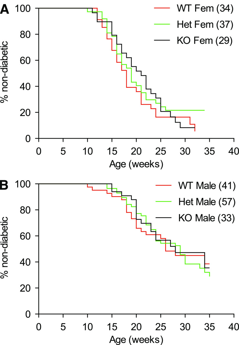
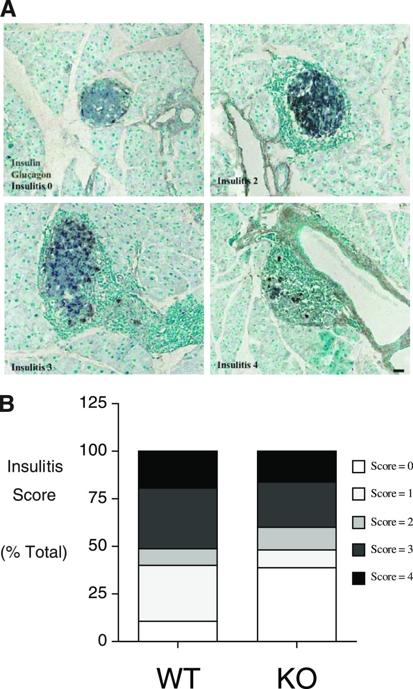
Results: The absence of G6pc2 did not affect the time of onset, incidence, or sex bias of type 1 diabetes in NOD/ShiLtJ mice. Insulitis was prominent in both groups, but whereas NOD/ShiLtJ G6pc2(+/+) islets contained CD8(+) T cells reactive to the G6pc2 NRP peptide, G6pc2 NRP-reactive T cells were absent in NOD/ShiLtJ G6pc2(-/-) islets.

Conclusions: These results demonstrate that G6pc2 is an important driver for the selection and expansion of islet-reactive CD8(+) T cells infiltrating NOD/ShiLtJ islets. However, autoreactivity to G6pc2 is not essential for the emergence of autoimmune diabetes. The results remain consistent with previous studies indicating that insulin may be the primary autoimmune target, at least in NOD/ShiLtJ mice.


Figures

FIG. 1.
FIG. 1.
Comparison of the incidence and time of onset of type 1 diabetes in wild-type NOD/ShiLtJ and NOD/ShiLtJ G6pc2−/− mice: the effect of sex. The incidence and time of onset of type 1 diabetes in male and female wild-type NOD/ShiLtJ, NOD/ShiLtJ G6pc2−/+, and NOD/ShiLtJ G6pc2−/− mice was compared starting at the age of 10 weeks. Clear differences were observed between male and female wild-type NOD/ShiLtJ mice (A), between male and female NOD/ShiLtJ G6pc2−/− mice (B), and between male and female NOD/ShiLtJ G6pc2−/+ mice (C). The number of animals studied is indicated in parentheses. Statistical significance between curves was determined by the log-rank (Mantel-Cox) test. Het, heterozygous; KO, knockout; WT, wild-type.
FIG. 2.
FIG. 2.
Comparison of the incidence and time of onset of type 1 diabetes in wild-type NOD/ShiLtJ and NOD/ShiLtJ G6pc2−/− mice: the effect of G6pc2 gene deletion. The cumulative incidence of type 1 diabetes in male and female wild-type NOD/ShiLtJ, NOD/ShiLtJ G6pc2−/+, and NOD/ShiLtJ G6pc2−/− mice was compared starting at the age of 10 weeks. No differences were observed between female wild-type, NOD/ShiLtJ G6pc2−/+, and NOD/ShiLtJ G6pc2−/− mice (A) or between male wild-type, NOD/ShiLtJ G6pc2−/+, and NOD/ShiLtJ G6pc2−/− mice (B). The number of animals studied is indicated in parentheses. Statistical significance between curves was determined by the log-rank (Mantel-Cox) test. Het, heterozygous; KO, knockout; WT, wild-type.
FIG. 3.
FIG. 3.
Insulitis in normoglycemic wild-type and G6pc2-deficient NOD/ShiLtJ mice. Paraffin sections of pancreata removed from 25-week-old normoglycemic male animals were stained by immunoperoxidase methods for the presence of insulin (gray) and glucagon (brown). Varying degrees of peri-islet mononuclear cell infiltration were evident, as visualized by the methyl green counter stain. Representative images (scale bar = 50 μm) are shown in A, whereas insulitis scores from five wild-type and five NOD/ShiLtJ G6pc2−/− mice are shown in B, based on the examination of a total of 127 wild-type and 151 G6pc2−/− islets. KO, knockout; WT, wild-type. (A high-quality digital representation of this figure is available in the online issue.)
FIG. 4.
FIG. 4.
Islet autoantigen–specific CD8+ T-cell responses in wild-type and G6pc2-deficient NOD/ShiLtJ mice. A and B: Islets were isolated from individual female wild-type or G6pc2−/− NOD/ShiLtJ mice at 12 weeks of age and cultured in the presence of interleukin-2 for 9 days. Cells were then stained with FITC-labeled anti-CD8α and PE-labeled MHC–class I tetramers loaded with the indicated peptides and analyzed by flow cytometry. Tetramers loaded with Flu-NP366–374 or Flu-NP147–155 were used as a negative control for tetramer staining. Data were electronically gated for CD8α-expressing cells. A: Representative flow cytometry data for one wild-type mouse and one G6pc2-deficient mouse. Numbers indicate the percentage of cells within the gated area. B: Summary of the flow cytometry data for four wild-type and four G6pc2-deficient mice from two experiments. C: Splenic and lymph-node cells were prepared from female wild-type or G6pc2−/− NOD/ShiLtJ mice at 12 weeks of age, and the frequency of NRP-V7–specific CD8+ T cells was determined by tetramer staining. Data were electronically gated for CD8α-expressing cells. Representative flow cytometry data are shown. D: Wild-type or G6pc2−/− NOD/ShiLtJ female mice were immunized with NRP-V7 peptide (100 μg per mouse) emulsified in complete Freund’s adjuvant. Two weeks later, splenic and draining lymph-node cells were prepared and stained with anti–CD8α-FITC and NRP-V7 or control Flu-NP tetramer and analyzed by flow cytometry. Data were electronically gated for CD8α-expressing cells. Representative flow cytometry data are shown. NS, not significant; WT, wild-type. (A high-quality color representation of this figure is available in the online issue.)

References

    1. Hutton JC, O’Brien RM. Glucose-6-phosphatase catalytic subunit gene family. J Biol Chem 2009;284:29241–29245 - PMC - PubMed
    1. Wang Y, Martin CC, Oeser JK, et al. . Deletion of the gene encoding the islet-specific glucose-6-phosphatase catalytic subunit-related protein autoantigen results in a mild metabolic phenotype. Diabetologia 2007;50:774–778 - PubMed
    1. Bouatia-Naji N, Rocheleau G, Van Lommel L, et al. . A polymorphism within the G6PC2 gene is associated with fasting plasma glucose levels. Science 2008;320:1085–1088 - PubMed
    1. Chen WM, Erdos MR, Jackson AU, et al. . Variations in the G6PC2/ABCB11 genomic region are associated with fasting glucose levels. J Clin Invest 2008;118:2620–2628 - PMC - PubMed
    1. Lieberman SM, Evans AM, Han B, et al. . Identification of the beta cell antigen targeted by a prevalent population of pathogenic CD8+ T cells in autoimmune diabetes. Proc Natl Acad Sci USA 2003;100:8384–8388 - PMC - PubMed

Publication types

MeSH terms