. 2011;117(2):98-105.
doi: 10.1254/jphs.11065fp. Epub 2011 Sep 7.

Effect of efonidipine on TGF-β1-induced cardiac fibrosis through Smad2-dependent pathway in rat cardiac fibroblasts

Affiliations

Effect of efonidipine on TGF-β1-induced cardiac fibrosis through Smad2-dependent pathway in rat cardiac fibroblasts

Bai Lei et al. J Pharmacol Sci. 2011.

Abstract

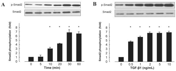
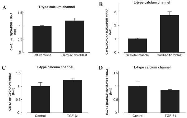
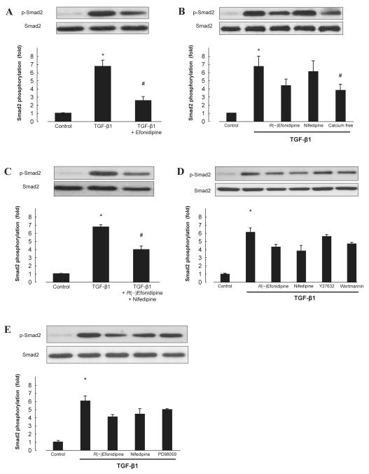
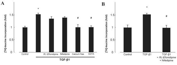
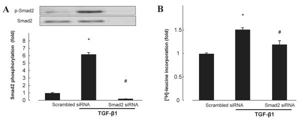
Transforming growth factor beta-1 (TGF-β1) plays a critical role in progression of cardiac fibrosis, which may involve intracellular calcium change. We examined effects of efonidipine, a dual T-type and L-type calcium channel blocker (CCB), on TGF-β1-induced fibrotic changes in neonatal rat cardiac fibroblast. T-type and L-type calcium channel mRNAs were highly expressed in cultured cardiac fibroblasts. TGF-β1 (5 ng/mL) significantly increased Smad2 phosphorylation and [(3)H]-leucine incorporation, which were attenuated by pretreatment with efonidipine (10 µM). Neither R(-)efonidipine (10 µM), selective T-type CCB, nor nifedipine (10 µM), selective L-type CCB, efficaciously inhibited both TGF-β1-induced Smad2 phosphorylation and [(3)H]-leucine incorporation. However, both were markedly attenuated by combination of R(-)efonidipine and nifedipine, EDTA, or calcium-free medium. Pretreatment with Smad2 siRNA significantly attenuated [(3)H]-leucine incorporation induced by TGF-β1. These data suggest that efonidipine elicits inhibitory effects on TGF-β1- and Smad2-dependent protein synthesis through both T-type and L-type calcium channel-blocking actions in cardiac fibroblasts.


Figures

Fig. 1
Fig. 1
Time-course and dose-dependent effect of TGF-β1 on Smad2 phosphorylation in cardiac fibroblasts. A: TGF-β1 (5 ng/mL) used to stimulate cells. B: Cells stimulated with TGF-β1 for 30 min. Bar graphs represent the mean ± S.E.M. (n = 6), expressed as the fold change in phosphorylation compared with unstimulated cells. *P < 0.05 vs. control cardiac fibroblasts.
Fig. 2
Fig. 2
Expression of T-type and L-type calcium channel mRNA in cardiac fibroblasts. A: A subunit of the T-type calcium channel, Cav3.1 (α1G), was highly expressed in cardiac fibroblasts. B: A subunit of the L-type calcium channel, Cav1.2 (CACNA1C), was highly expressed in cardiac fibroblasts. C and D: TGF-β1 (5 ng/mL, 24 h) did not increase T-type or L-type calcium channel in cardiac fibroblasts. Bar graphs represent the mean ± S.E.M. (n = 6), expressed as the fold change in mRNA expression compared to positive controls; mRNA levels are normalized to GAPDH mRNA levels.
Fig. 3
Fig. 3
Effect of CCBs on TGF-β1–induced Smad2 phosphorylation in cardiac fibroblasts. Smad2 phosphorylation induced by TGF-β1 was measured in the absence or presence of efonidipine (10 μM) (A); R(−)efonidipine (10 μM), nifedipine (10 μM), calcium-free medium (B); combination of R(−)efonidipine and nifedipine (C); Y27632 (1 μM), wortmannin (100 nM) (D); or PD98059 (30 μM) (E). Bar graphs represent the mean ± S.E.M. (n = 6), expressed as the fold change in phosphorylation compared with unstimulated cells. *P < 0.05, control cardiac fibroblasts vs. TGF-β1 alone; #P < 0.05, TGF-β1 alone vs. TGF-β1 with treatment.
Fig. 4
Fig. 4
Effect of TGF-β1 on [3H]-leucine incorporation in the absence or presence of R(−)efonidipine, nifedipine, calcium-free medium, EDTA (A) or the combination of R(−)efonidipine and nifedipine (B). Bar graphs represent the mean ± S.E.M. (n = 6), expressed as the fold change compared with unstimulated cells. *P < 0.05, control cardiac fibroblasts vs. TGF-β1 alone; #P < 0.05, TGF-β1 alone vs. TGF-β1 with treatment.
Fig. 5
Fig. 5
Effect of Smad2 siRNA on protein expression (A) and TGF-β1–induced [3H]-leucine incorporation (B) in cardiac fibroblasts. Bar graphs represent the mean ± S.E.M. (n = 6), expressed as the fold change compared with unstimulated cells. *P < 0.05, control cardiac fibroblasts vs. TGF-β1 alone; #P < 0.05, TGF-β1 alone vs. TGF-β1 with treatment.

Similar articles

Cited by

References

    1. Akiyama-Uchida Y, Ashizawa N, Ohtsuru A, Seto S, Tsukazaki T, Kikuchi H, et al. Norepinephrine enhances fibrosis mediated by TGF-beta in cardiac fibroblasts. Hypertension. 2002;40:148–154. - PubMed
    1. Takeda N, Manabe I, Uchino Y, Eguchi K, Matsumoto S, Nishimura S, et al. Cardiac fibroblasts are essential for the adaptive response of the murine heart to pressure overload. J Clin Invest. 2010;120:254–265. - PMC - PubMed
    1. Nagamatsu Y, Nishida M, Onohara N, Fukutomi M, Maruyama Y, Kobayashi H, et al. Heterotrimeric G protein G alpha13-induced induction of cytokine mrnas through two distinct pathways in cardiac fibroblasts. J Pharmacol Sci. 2006;101:144–150. - PubMed
    1. Eghbali M, Tomek R, Sukhatme VP, Woods C, Bhambi B. Differential effects of transforming growth factor-beta 1 and phorbol myristate acetate on cardiac fibroblasts. Regulation of fibrillar collagen mrnas and expression of early transcription factors. Circ Res. 1991;69:483–490. - PubMed
    1. Sigel AV, Centrella M, Eghbali-Webb M. Regulation of proliferative response of cardiac fibroblasts by transforming growth factor-beta 1. J Mol Cell Cardiol. 1996;28:1921–1929. - PubMed

Publication types

MeSH terms