

Molecular genetics of retinal degeneration: A Drosophila perspective
- PMID: 21897116
- PMCID: PMC3266078
- DOI: 10.4161/fly.5.4.17809
Molecular genetics of retinal degeneration: A Drosophila perspective
Abstract
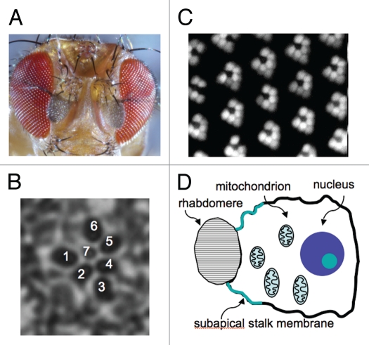
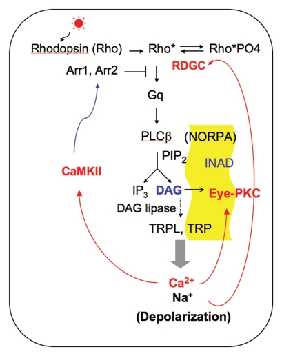
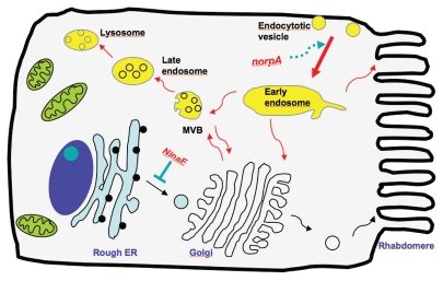
Inherited retinal degeneration in Drosophila has been explored for insights into similar processes in humans. Based on the mechanisms, I divide these mutations in Drosophila into three classes. The first consists of genes that control the specialization of photoreceptor cells including the morphogenesis of visual organelles (rhabdomeres) that house the visual signaling proteins. The second class contains genes that regulate the activity or level of the major rhodopsin, Rh1, which is the light sensor and also provides a structural role for the maintenance of rhabdomeres. Some mutations in Rh1 (NinaE) are dominant due to constitutive activity or folding defects, like autosomal dominant retinitis pigmentosa (ADRP) in humans. The third class consists of genes that control the Ca ( 2+) influx directly or indirectly by promoting the turnover of the second messenger and regeneration of PIP 2, or mediate the Ca ( 2+) -dependent regulation of the visual response. These gene products are critical for the increase in cytosolic Ca ( 2+ ) following light stimulation to initiate negative regulatory events. Here I will focus on the signaling mechanisms underlying the degeneration in norpA, and in ADRP-type NinaE mutants that produce misfolded Rh1. Accumulation of misfolded Rh1 in the ER triggers the unfolded protein response (UPR), while endosomal accumulation of activated Rh1 may initiate autophagy in norpA. Both autophagy and the UPR are beneficial for relieving defective endosomal trafficking and the ER stress, respectively. However, when photoreceptors fail to cope with the persistence of these stresses, a cell death program is activated leading to retinal degeneration.
Figures
References
-
- Franceschini N. Pupil and psueopupil in the compound eye of Drosophila. In: Wehner R, editor. Information Processing in the Visual Systems of Arthropods. Berlin: Springer-Verlag; 1972. pp. 75–82.
-
- Knust E. Photoreceptor morphogenesis and retinal degeneration: lessons from Drosophila. Curr Opin Neurobiol. 2007;17:541–547. - PubMed
-
- Brand AH, Perrimon N. Targeted gene expression as a means of altering cell fates and generating dominant phenotypes. Development. 1993;118:401–415. - PubMed
-
- Elliott DA, Brand AH. The GAL4 system: a versatile system for the expression of genes. Methods Mol Biol. 2008;420:79–95. - PubMed
-
- Carthew RW. Gene silencing by double-stranded RNA. Curr Opin Cell Biol. 2001;13:244–248. - PubMed
Publication types
MeSH terms
Substances
Grants and funding
LinkOut - more resources
Full Text Sources
Molecular Biology Databases
Research Materials