. 2011 Oct 11;105(8):1210-7.
doi: 10.1038/bjc.2011.348. Epub 2011 Sep 6.

Activin A inhibits vascular endothelial cell growth and suppresses tumour angiogenesis in gastric cancer

Affiliations

Activin A inhibits vascular endothelial cell growth and suppresses tumour angiogenesis in gastric cancer

H Kaneda et al. Br J Cancer. .

Abstract

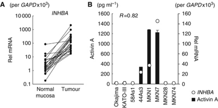
Background: Activin A is a multi-functional cytokine belonging to the transforming growth factor-β (TGF-β) superfamily; however, the effect of activin A on angiogenesis remains largely unclear. We found that inhibin β A subunit (INHBA) mRNA is overexpressed in gastric cancer (GC) specimens and investigated the effect of activin A, a homodimer of INHBA, on angiogenesis in GC.

Methods: Anti-angiogenic effects of activin A via p21 induction were evaluated using human umbilical vein endothelial cells (HUVECs) in vitro and a stable INHBA-introduced GC cell line in vivo.

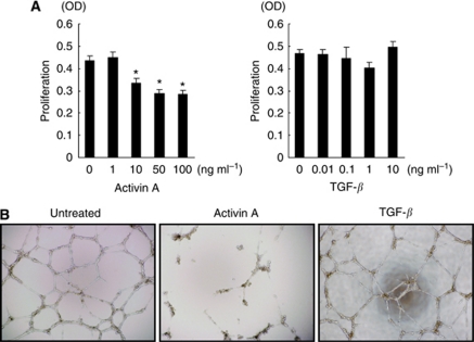
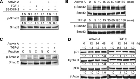
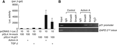
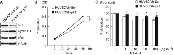
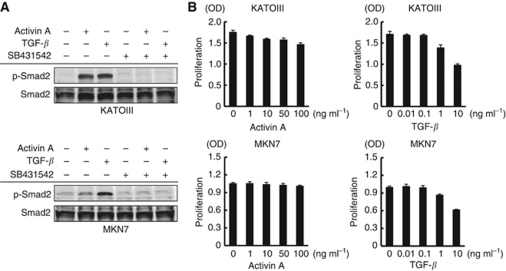
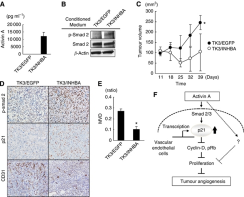
Results: Compared with TGF-β, activin A potently inhibited the cellular proliferation and tube formation of HUVECs with induction of p21. A promoter assay and a chromatin immunoprecipitation assay revealed that activin A directly regulates p21 transcriptional activity through Smads. Stable p21-knockdown significantly enhanced the cellular proliferation of HUVECs. Notably, stable p21-knockdown exhibited a resistance to activin-mediated growth inhibition in HUVECs, indicating that p21 induction has a key role on activin A-mediated growth inhibition in vascular endothelial cells. Finally, a stable INHBA-introduced GC cell line exhibited a decrease in tumour growth and angiogenesis in vivo.

Conclusion: Our findings highlight the suppressive role of activin A, unlike TGF-β, on tumour growth and angiogenesis in GC.


Figures

Figure 1
Figure 1
Overexpression of INHBA (inhibin β A) mRNA in GC specimens and secretion of its homodimer form, activin A, in GC cell lines. (A) The mRNA expressions of INHBA in 24 GC and paired non-cancerous gastric mucosa samples were determined using real-time RT–PCR. (B) A strong correlation between the expressions of INHBA mRNA and activin A protein was observed in GC cell lines, as determined using real-time RT–PCR and an ELISA, respectively. Rel mRNA: normalised mRNA expression levels (INHBA/GAPD × 103). The correlation coefficient is shown in the figure.
Figure 2
Figure 2
Activin A potently inhibits the proliferation and tube formation of HUVECs. (A) HUVECs were cultured in 96-well plates and stimulated with the indicated doses of activin A or TGF-β for 72 h. The cell proliferation was assayed using an MTT assay. Columns: mean of independent triplicate experiments. Bars: s.d. *P<0.05. (B) Effect of activin A on tube formation in HUVECs. HUVECs were cultured with normal medium (untreated) or activin A (10 ng ml–1) or TGF-β (1 ng ml–1) containing medium for 48 h and the cells were seeded in 96-well culture plates (1.5 × 10⁴ cells per well) precoated with 80 μl Matrigel and cultured with normal medium (untreated) or activin A (10 ng ml–1) or TGF-β (1 ng ml–1). After 16 h of incubation, the wells were photographed using a microscope.
Figure 3
Figure 3
Activin A mediates the persistent phosphorylation of Smad2 and p21 induction in HUVECs. (A) HUVECs were treated with or without 2 μM of SB341542 for 30 min, then stimulated with activin A (10 ng ml–1) or TGF-β (1 ng ml–1) for 1 h. The phosphorylation and expression levels of Smad2 were evaluated using western blot. (B) Time-course analysis with activin A or TGF-β-induced Smad2 phosphorylation. HUVECs were stimulated with 10 ng ml–1 activin A or 1 ng ml–1 TGF-β for the indicated time periods. (C) Activin A or TGF-β-induced nuclear translocation of Smad2. HUVECs were stimulated with or without activin A (10 ng ml–1) or TGF-β (1 ng ml–1) for 1 h. Nuclear and cytosolic protein fractions were then analysed using a western blot analysis. C= cytosolic fraction; N=nuclear fraction. (D) Expression changes of cell cycle-related proteins by stimulation with activin A (10 ng ml–1) or TGF-β (1 ng ml–1) for the indicated time period in HUVECs. A western blot analysis was performed using anti-p21, cyclin D, and phosphorylated Rb antibodies. β-Actin was used as an internal control.
Figure 4
Figure 4
Activin A directly increases p21 promoter activity. (A) p21 promoter activity was determined using a luciferase assay. HEK293 cells were transiently transfected with luciferase vectors containing empty or p21 promoters (pGL4.14-mock or pGL4.14-p21) and then stimulated with activin A (10 ng ml–1) or TGF-β (1 ng ml–1) for 24 h. The data were normalised by β-galactosidase activity of co-transfected with the Lac Z vector in at least three independent experiments. Columns: mean of experiments. Bars: s.d. (B) ChIP of activin A-induced Smads on the promoter of p21. HUVECs were treated with 10 ng ml–1 of activin A for 1 h and collected for analysis. The data show the PCR amplification of the p21 promoter using inputs (1% of chromatin used for ChIP) or ChIPs using smad3 or smad4 antibodies as templates. Primers to the GAPDH promoter were used as a negative control. IgG: non-specific IgG as a control.
Figure 5
Figure 5
p21 induction has a key role in activin A-mediated growth inhibition in vascular endothelial cells. (A) Stable HUVECs transfected with control or p21-knockdown viral shRNA vector (HUVEC/sh-Scr and HUVEC/sh-p21) were evaluated using a western blot analysis using p21, cyclin D, and phospho-Rb antibodies. β-Actin was used as an internal control. (B) The cell growth curves of HUVEC/sh-Scr and HUVEC/sh-p21 cells were evaluated using an MTT assay in triplicate experiments. *P<0.05. (C) Growth inhibition of HUVEC/sh-Scr and HUVEC/sh-p21 cells by activin A stimulation. The cells were stimulated with the indicated doses of activin A for 48 h. Cell growth was determined using an MTT assay. Data were shown as the percentage of activin A-untreated controls and are shown as the mean±s.d. of at least three independent experiments. *P<0.05.
Figure 6
Figure 6
Activin A inhibits the cellular proliferation of KATOIII cells but does not inhibit the proliferation of MKN7 cells. (A) GC cell lines KATOIII and MKN7 were treated with or without 2 μM of SB341542 for 30 min, then stimulated with activin A (10 ng ml–1) or TGF-β (1 ng ml–1) for 1 h. The phosphorylation and expression levels of Smad2 were evaluated using a western blot. (B) KATOIII and MKN7 cells were cultured in 96-well plates and stimulated with the indicated doses of activin A or TGF-β for 72 h. Cellular proliferation was assayed using an MTT assay. Columns: mean of independent triplicate experiments. Bars: s.d.
Figure 7
Figure 7
Overexpression of INHBA potently inhibited tumour growth and angiogenesis in GC in vivo. (A) TU-KATOIII GC cells were stably transfected with an INHBA (TK3/INHBA cells) or control vector (TK3/EGFP cells). Activin A secretion in the conditioned medium was analysed using an ELISA. (B) Activity of exogenous activin A on phosphorylation of smad2. Conditioned media from TK3/INHBA or /EGFP cells were exposed to HUVECs for 1 h and the phosphorylation of Smad2 in the HUVECs was assessed using a western blot analysis. β-Actin was used as an internal control. (C) Effect of overexpression of INHBA on tumour growth. TK3/EGFP and TK3/INHBA cells (1 × 10⁷ cells) were subcutaneously inoculated into mice and evaluated for tumour growth in vivo. The data indicate the mean±s.d. *P<0.05. (D and E) CD31 staining for tumour specimens. Microvessel density (MVD) was evaluated using CD31-positive endothelial cells in tumour specimens using a computer-assisted image analysis. *P<0.05. (F) Diagram of the proposed mechanism of activin A on vascular endothelial cells and in angiogenesis.

Similar articles

Cited by

References

    1. Burdette JE, Jeruss JS, Kurley SJ, Lee EJ, Woodruff TK (2005) Activin A mediates growth inhibition and cell cycle arrest through Smads in human breast cancer cells. Cancer Res 65: 7968–7975 - PubMed
    1. Chen YG, Lui HM, Lin SL, Lee JM, Ying SY (2002) Regulation of cell proliferation, apoptosis, and carcinogenesis by activin. Exp Biol Med (Maywood) 227: 75–87 - PubMed
    1. Datto MB, Li Y, Panus JF, Howe DJ, Xiong Y, Wang XF (1995) Transforming growth factor beta induces the cyclin-dependent kinase inhibitor p21 through a p53-independent mechanism. Proc Natl Acad Sci USA 92: 5545–5549 - PMC - PubMed
    1. Dawid IB, Taira M, Good PJ, Rebagliati MR (1992) The role of growth factors in embryonic induction in Xenopus laevis. Mol Reprod Dev 32: 136–144 - PubMed
    1. el-Deiry WS, Tokino T, Velculescu VE, Levy DB, Parsons R, Trent JM, Lin D, Mercer WE, Kinzler KW, Vogelstein B (1993) WAF1, a potential mediator of p53 tumor suppression. Cell 75: 817–825 - PubMed

Publication types

MeSH terms