Review
. 2011 Sep 7;12(10):585-601.
doi: 10.1038/nrn3085.

A pathophysiological framework of hippocampal dysfunction in ageing and disease

Affiliations
Review

A pathophysiological framework of hippocampal dysfunction in ageing and disease

Scott A Small et al. Nat Rev Neurosci. .

Abstract

The hippocampal formation has been implicated in a growing number of disorders, from Alzheimer's disease and cognitive ageing to schizophrenia and depression. How can the hippocampal formation, a complex circuit that spans the temporal lobes, be involved in a range of such phenotypically diverse and mechanistically distinct disorders? Recent neuroimaging findings indicate that these disorders differentially target distinct subregions of the hippocampal circuit. In addition, some disorders are associated with hippocampal hypometabolism, whereas others show evidence of hypermetabolism. Interpreted in the context of the functional and molecular organization of the hippocampal circuit, these observations give rise to a unified pathophysiological framework of hippocampal dysfunction.


Figures
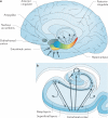
Figure 1
Figure 1. The organization of the hippocampal formation
a | The hippocampal formation, which is made up of the entorhinal cortex and hippocampus (shown in colour) extends over the anterior-to-posterior axis of the brain. The colour gradients reflect the topological input–output relations between the hippocampal formation and other brain areas, as well as its internal functional and molecular organization. Input to the hippocampus is shown by solid arrows, hippocampal outflow is shown by dashed arrows. Cortical and subcortical information funnels onto superficial layers of the entorhinal cortex, and this input is organized in an anterior–medial to posterior–lateral gradient (shown by the colour gradient in the entorhinal cortex). This anatomical gradient is largely preserved as the entorhinal cortex conveys this information to the hippocampus (shown by the corresponding colour code in the hippocampus). The long-axis gradient is preserved in the output pattern of the hippocampus. As well as reconnecting with the entorhinal cortex, the hippocampus monosynaptically connects with — from anterior to posterior — the orbitofrontal cortex, anterior cingulate, amygdala, nucleus accumbens and posterior cingulate. b | In the hippocampal transverse axis, superficial layers of the entorhinal cortex connect with the dentate gyrus (DG), CA3, CA1 and the subiculum (Sub). The trisynaptic circuit connects the dentate gyrus to CA3, to CA1 and to the subiculum. Through auto-association fibres, CA3 neurons interconnect with other CA3 neurons throughout the long axis. The CA1 and primarily the subiculum provide the main hippocampal outflow (shown by dashed arrows), back to the deep layers of the entorhinal cortex and also to a range of cortical and subcortical sites (as shown in part a).
Figure 2
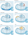
Figure 2. Regional vulnerability and metabolic state differentiate disorders that affect the hippocampal formation
Although multiple hippocampal subregions can be affected in disorders, by comparing patterns of alterations that are observed by functional and structural MRI it is possible to isolate individual subregions differentially affected by each disorder. Furthermore, functional imaging techniques that are sensitive to metabolic state have suggested that some hippocampus-based disorders are characterized by hypometabolism (shown in blue), whereas others are abnormally hypermetabolic (shown in red). a | Alzheimer's disease, vascular disease and ageing all contribute to hippocampal alterations in late life. A direct comparison suggests that the entorhinal cortex (EC) is differentially associated with Alzheimer's disease and the CA1 with vascular disease, whereas the ageing process per se seems to differentially target the dentate gyrus (DG). Hypometabolism has been localized to the EC in Alzheimer's disease, to CA1 in vascular disease and the DG in ageing. b | Schizophrenia and depression typically begin during adolescence, and post-traumatic stress disorder (PTSD) currently has its highest incidence in young adulthood. A direct comparison suggests that CA1 is differentially associated with schizophrenia and that the subiculum (Sub) may be differentially associated with depression. In the case of PTSD, both CA3 and the DG have been implicated. Evidence for global hippocampal hypermetabolism exists for both schizophrenia and depression. In schizophrenia, this hypermetabolism may be driven by hypermetabolism in CA1, whereas subicular hypermetabolism in depression is a hypothesis that remains untested (shown in yellow). In PTSD the metabolic state of the hippocampal formation is as yet undetermined (shown in yellow).
Figure 3
Figure 3. A proposed ‘functional map’ of the hippocampal circuit
Although multiple subregions are engaged by different tasks, as neural information flows through the hippocampal circuit, each hippocampal subregion is thought to perform a distinct cognitive or computational operation. It is possible to begin to chart a ‘functional map’ of the hippocampal circuit, linking distinct operations to individual hippocampal subregions. Here, CA1 is important in integration of inputs, the dentate gyrus (DG) has a role in pattern separation, CA3 has a role in pattern completion, the subiculum (Sub) is involved in memory retrieval and the entorhinal cortex (EC) is involved in brief retention in hippocampus-dependent memory tasks.
Figure 4
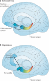
Figure 4. Proposed hippocampus-based networks in schizophrenia and depression
a | In schizophrenia, hypermetabolism in the anterior hippocampus occurs early in the disease process, and through monosynaptic connections this can be linked to orbitofrontal hypermetabolism that emerges later in the disease course. In animal models, hyperactivity in the anterior hippocampus stimulates the nucleus accumbens, leading to increased striatal dopamine release. b | Studies in patients with depression suggest that hypermetabolism in the anterior hippocampus occurs early in the disease process, and via monosynaptic connections this may be linked to hypermetabolism observed in the amygdala and the anterior cingulate.

Comment in

References

    1. Zola-Morgan S, Squire LR, Amaral DG. Human amnesia and the medial temporal region: enduring memory impairment following a bilateral lesion limited to field CA1 of the hippocampus. J. Neurosci. 1986;6:2950–2967. - PMC - PubMed
    1. Wu W, et al. The brain in the age of old: the hippocampal formation is targeted differentially by diseases of late life. Ann. Neurol. 2008;64:698–706. - PMC - PubMed
    1. Sloviter RS, Sollas AL, Dean E, Neubort S. Adrenalectomy-induced granule cell degeneration in the rat hippocampal dentate gyrus: characterization of an in vivo model of controlled neuronal death. J. Comp. Neurol. 1993;330:324–336. - PubMed
    1. Fanselow MS, Dong HW. Are the dorsal and ventral hippocampus functionally distinct structures? Neuron. 2010;65:7–19. - PMC - PubMed
    1. Zhao X, et al. Transcriptional profiling reveals strict boundaries between hippocampal subregions. J. Comp. Neurol. 2001;441:187–196. - PubMed

Publication types