

Giant primary ossified cavernous hemangioma of the skull in an adult: A rare calvarial tumor
- PMID: 21897684
- PMCID: PMC3159357
- DOI: 10.4103/0976-3147.83587
Giant primary ossified cavernous hemangioma of the skull in an adult: A rare calvarial tumor
Abstract
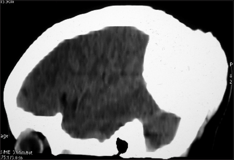
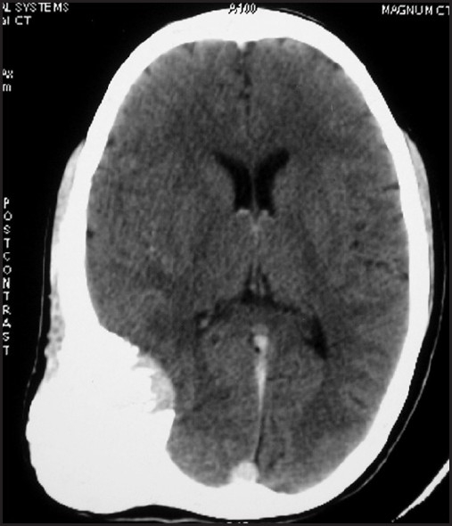
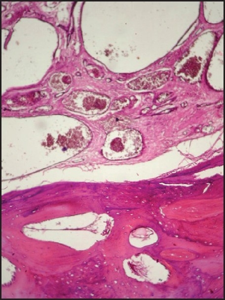
Primary intraosseous cavernous hemangiomas (PICHs) of the cranium are rare benign vascular tumors that account for about 0.2 % of all bone tumors and 10 % of benign skull tumors. They generally present as osteolytic lesions with honeycomb pattern of calcification. Completely ossified cavernous hemangioma of the calvarium in an adult has not been reported previously. A 28-year-old female presented to us with a large right parietal skull mass that had been present since the last 15 years. Total resection of the lesion was performed. Pathological examination was suggestive of cavernous hemangioma of the skull bone. Cavernous hemangioma should be considered as one of the differential diagnosis in any case of bony swelling of the calvarium so that adequate preoperative planning can be made to minimize blood loss and subsequent morbidity.
Keywords: Adult; cavernous hemangioma; ossified calvarial mass; skull lesion.
Conflict of interest statement
Figures
References
-
- Wyke BD. Primary hemangioma of skull: A rare cranial tumour. Am J Roentgenol. 1946;61:302–16. - PubMed
-
- Naama O, Gazzaz M, Akhaddar A, Belhachmi A, Asri A, Elmostarchid B, et al. Cavernous hemangioma of the skull: 3 case reports. Surg Neurol. 2008;70:654–9. - PubMed
-
- Ajja A, Oukacha N, Gazzaz M, Akhaddar A, Elmostarchid B, Kadiri B, et al. Cavernous hemangioma of the parietal bone: A case report. J Neurosurg Sci. 2005;49:159–62. - PubMed
-
- Hombal AG, Hegde KK. Giant Haemangioma of the Scalp: A Case Report. Ind J Radiol Imag. 2006;16:41–3.
Publication types
LinkOut - more resources
Full Text Sources