Pancreatobiliary reflux resulting in pancreatic ascites and choleperitoneum after gallbladder perforation
- PMID: 21897795
- PMCID: PMC3166807
- DOI: 10.1159/000161567
Pancreatobiliary reflux resulting in pancreatic ascites and choleperitoneum after gallbladder perforation
Abstract
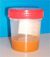
A 65-year-old man with chronic hepatitis C and no history of alcohol abuse was admitted to our liver unit for the recent development of massive ascites and presumed hepatorenal syndrome. In the preceding two weeks, he had received medical treatment for acute pancreatitis and cholecystitis. Abdominal paracentesis demonstrated a cloudy, orange peritoneal fluid, with total protein concentration 3.6 g/dl, serum-ascites albumin gradient 1.0 g/dl, and ratios of ascites-serum bilirubin and amylase approximately 8:1. Diagnostic imaging demonstrated no pancreatic pseudocysts. Ten days later, at laparotomy, acalculous perforation of the gallbladder was identified. After cholecystectomy, amylase concentration in the ascitic fluid dropped within a few days to 40% of serum values; ascites disappeared within a few weeks. We conclude that in the presence of a perforated gallbladder, pancreatobiliary reflux was responsible for this unusual combination of choleperitoneum and pancreatic ascites, which we propose to call pancreatobiliary ascites.
Keywords: Ascites; Cholecystitis; Pancreatitis, acute necrotizing; Peritonitis.
Figures
Similar articles
-
Ascitic fluid bilirubin concentration as a key to choleperitoneum.J Clin Gastroenterol. 1987 Oct;9(5):543-5. doi: 10.1097/00004836-198710000-00011. J Clin Gastroenterol. 1987. PMID: 3680904
-
Black ascitic fluid in a patient with history of alcohol abuse: report of an unusual case and literature review.Oxf Med Case Reports. 2020 May 23;2020(4):omaa023. doi: 10.1093/omcr/omaa023. eCollection 2020 Apr. Oxf Med Case Reports. 2020. PMID: 32477573 Free PMC article.
-
Pancreatic ascites managed with a conservative approach: a case report.J Med Case Rep. 2020 Sep 15;14(1):154. doi: 10.1186/s13256-020-02463-0. J Med Case Rep. 2020. PMID: 32928282 Free PMC article.
-
Does this patient have bacterial peritonitis or portal hypertension? How do I perform a paracentesis and analyze the results?JAMA. 2008 Mar 12;299(10):1166-78. doi: 10.1001/jama.299.10.1166. JAMA. 2008. PMID: 18334692 Review.
-
Pancreatic ascites: update on diagnosis and management.Ann Gastroenterol. 2025 May-Jun;38(3):247-254. doi: 10.20524/aog.2025.0961. Epub 2025 Apr 22. Ann Gastroenterol. 2025. PMID: 40371206 Free PMC article. Review.
Cited by
-
Idiopathic perforation of acalculous gallbladder after insertion of a transpapillary pancreatic stent.Endosc Int Open. 2016 Aug;4(8):E838-40. doi: 10.1055/s-0042-109598. Epub 2016 Aug 10. Endosc Int Open. 2016. PMID: 27540570 Free PMC article.
References
-
- Uchiyama T, Yamamoto T, Mizuta E, Suzuki T. Pancreatic ascites – a collected review of 37 cases in Japan. Hepatogastroenterology. 1989;36:244–248. - PubMed
-
- Fernandez-Cruz L, Margarona E, Llovera J, Lopez-Boado MA, Saenz H. Pancreatic ascites. Hepatogastroenterology. 1993;40:150–154. - PubMed
-
- Behar J, Corazziari E, Guelrud M, Hogan W, Sherman S, Toouli J. Functional gallbladder and sphincter of Oddi disorders. Gastroenterology. 2006;130:1498–1509. - PubMed
-
- Runyon BA. Ascitic fluid bilirubin concentration as a key to choleperitoneum. J Clin Gastroenterol. 1987;9:543–545. - PubMed
Publication types
LinkOut - more resources
Full Text Sources
Miscellaneous


