A rare case of synchronous adrenocortical carcinoma and renal cell carcinoma
- PMID: 21897901
- PMCID: PMC3156544
- DOI: 10.4103/2230-8210.83409
A rare case of synchronous adrenocortical carcinoma and renal cell carcinoma
Abstract
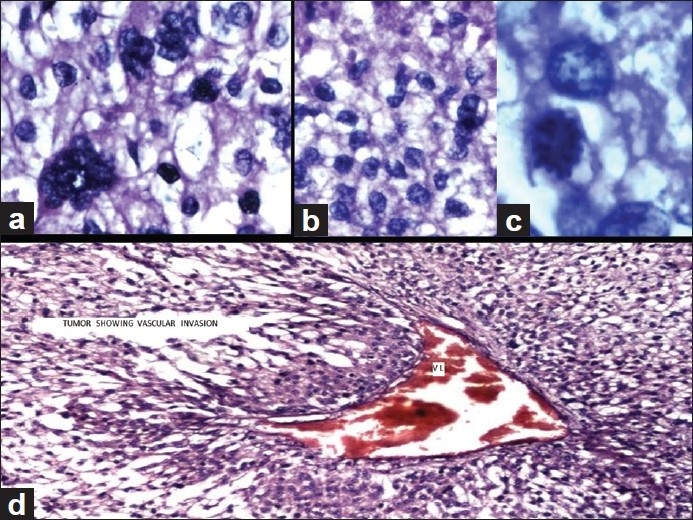
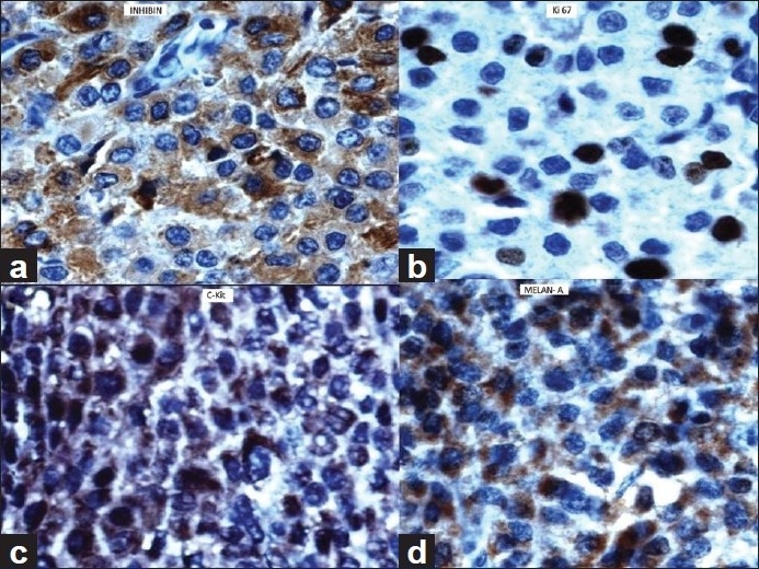
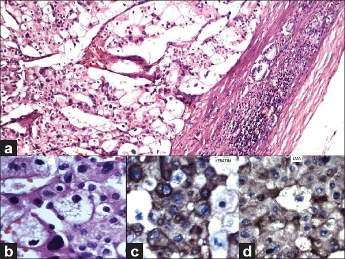
We present here a rare case of synchronous adrenocortical carcinoma (ACC) and renal cell carcinoma (RCC). A 27-year-old woman presented with gradual abdominal distension, hematuria, and loss of weight of 3-months duration. She gave a history of treatment for hypertension. The computed axial tomography (CT) scan revealed a large retroperitoneal mass. Her urinary VMA was slightly elevated. Her 24-h urinary metanephrine level was normal. The patient underwent left adrenalectomy, left nephrectomy, spleenectomy, and distal pancreactomy with segmental colonic resection. Postoperative pathology revealed ACC of left suprarenal measuring 22 × 19 × 18 cm(3) and RCC involving the left upper pole of kidney measuring 3 × 2 × 1 cm(3).
Keywords: Adrenalectomy; adrenocortical carcinoma; renal cell carcinoma.
Conflict of interest statement
Figures
References
-
- Wick MR, Cherwitz DL, McGlennen RC. Adrenocortical carcinoma.An immunohistochemical comparison with renal cell carcinoma. Am J Pathol. 1986;122:343–52. - PMC - PubMed
-
- Foucar E, Dehner LP. Renal cell carcinoma occurring with contralateral adrenal metastasis: A clinical and pathologic trap. Arch Surg. 1979;114:955–63. - PubMed
-
- Jani P, Nasr AL, Demellawy DE. Synchronous renal cell carcinoma and adrenocortical carcinoma: A rare case report and clinicopathologic approach. Can J Urol. 2008;15:4016–9. - PubMed