. 2012 Jan;55(1):86-97.
doi: 10.1002/hep.24629.

Immunization with aspartate-β-hydroxylase-loaded dendritic cells produces antitumor effects in a rat model of intrahepatic cholangiocarcinoma

Affiliations

Immunization with aspartate-β-hydroxylase-loaded dendritic cells produces antitumor effects in a rat model of intrahepatic cholangiocarcinoma

Takehiro Noda et al. Hepatology. 2012 Jan.

Abstract

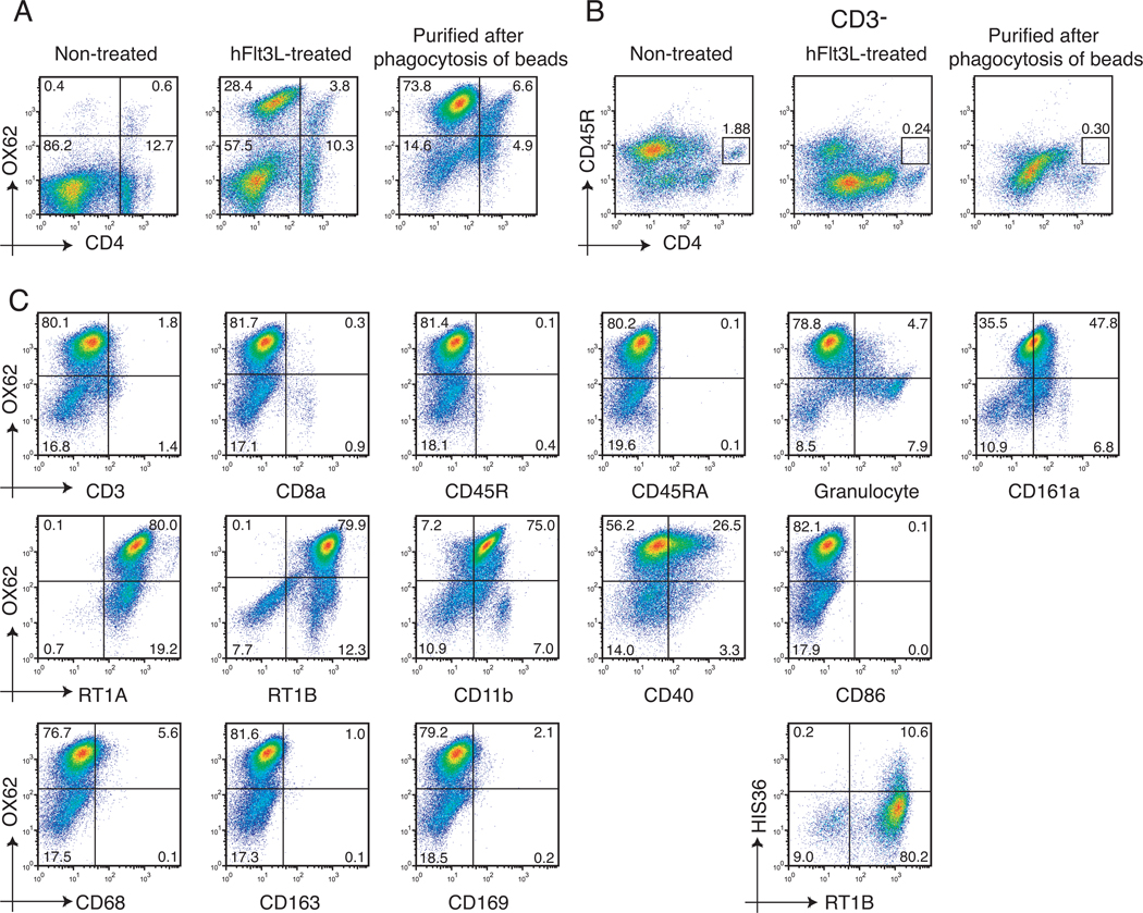
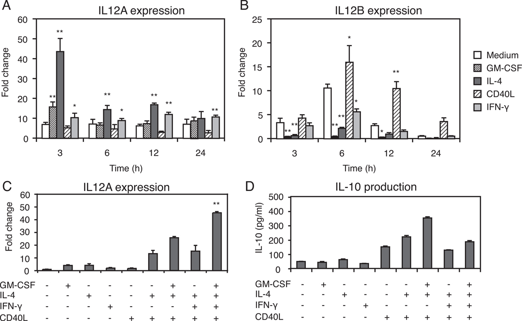
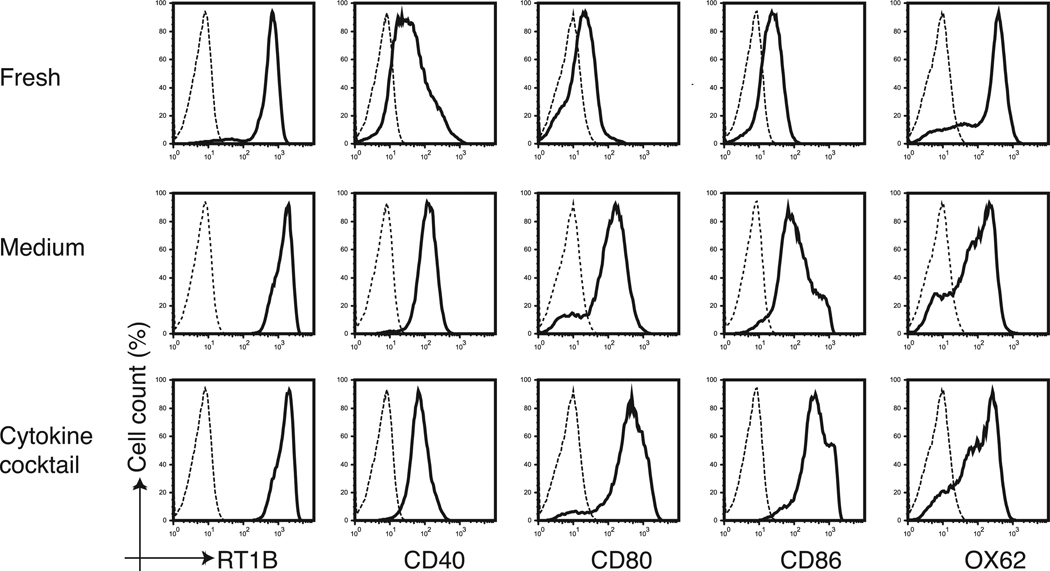
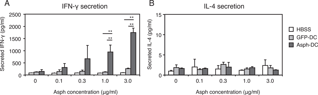
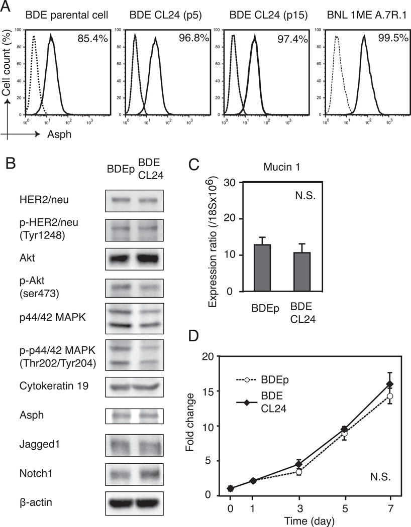
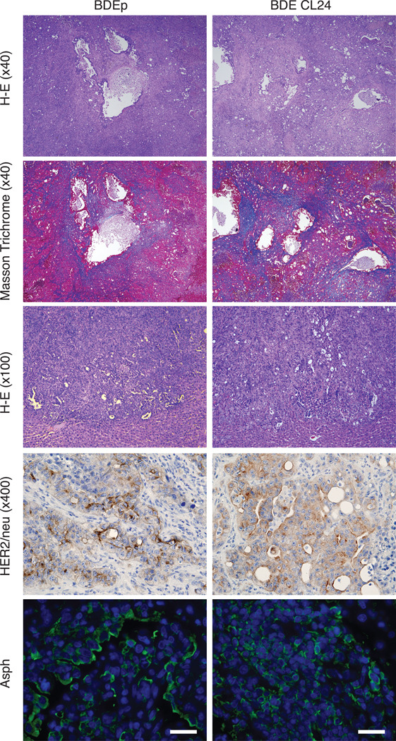
Dendritic cells (DCs) capture and process proteins and present peptides on the cell surface in the context of major histocompatibility complex I and II molecules to induce antigen-specific T cell immune responses. The aims of this study were to (1) employ an expanded and purified DC population and load them with aspartate-β-hydroxylase (ASPH), a highly expressed tumor-associated cell surface protein, and (2) to determine if immunization induced antitumor effects in an orthotopic rat model of intrahepatic cholangiocarcinoma. Splenocytes were incubated with ASPH-coated beads and passed through a magnetic field to yield an 80% pure DC OX62+ population. This DC subset was stimulated with granulocyte-macrophage colony-stimulating factor, interleukin-4, CD40L, and interferon-γ, resulting in a 40-fold increase in interleukin-12A messenger RNA expression to subsequently generate a T helper 1-type immune response. After incubation with the cytokine cocktail, DCs were found to have matured, as demonstrated by increased expression of CD40, CD80, and CD86 costimulatory molecules. Immunization with ASPH-loaded DCs induced antigen-specific immunity. A clone of the parental tumorigenic rat BDEneu cholangiocyte cell line, designated BDEneu-CL24, was found to have the highest number of cells expressing this surface protein (97%); it maintained the same phenotypic characteristics of the parental cell line and was used to produce intrahepatic tumors in immunocompetent syngeneic Fisher-344 rats. Immunization with ASPH-loaded DCs generated cytotoxicity against cholangiocarcinoma cells in vitro and significantly suppressed intrahepatic tumor growth and metastasis, and was associated with increased CD3+ lymphocyte infiltration into the tumors.

Conclusion: These findings suggest that immunization with ASPH-loaded DCs may constitute a novel therapeutic approach for intrahepatic cholangiocarcinoma, because this protein also appears to be highly conserved and expressed on human hepatobiliary tumors.


Conflict of interest statement

Conflict of interest: The authors have declared that no conflict of interest exists.

Figures

Figure 1
Figure 1. Flow cytometry analysis of cell surface markers on purified DCs
(A) The changes observed in the OX62+DCs population obtained from non-expanded (normal) expanded and purified from splenocyte populations. Cells are displayed in a OX62 and CD4 plot. (B) Detection of pDCs (CD3-CD4+CD45R+) in non-treated (normal), hFlt3L-treated splenocytes, or purified after phagocytosis of beads using flow cytometry. (C) The phenotype of the hFlt3L stimulated and purified DC population. To detect the HIS36+ cells, cells were stained with PE-conjugated anti-macrophage subset (HIS36) antibody and FITC-conjugated anti-RT1B antibody.
Figure 2
Figure 2. Cytokine expression in purified DCs
(A) Transient expression of IL12A mRNA of the IL-12p35 subunit in purified DCs stimulated for the indicated times with GM-CSF (4 ng/ml), IL-4 (100 ng/ml), or CD40L (1 µg/ml), or IFN-γ (20 ng/ml). (B) The expression of IL12B mRNA of the IL-12p40 subunit after cytokine stimulation. In panel (A) and (B), the expression of IL12A mRNA or IL12B mRNA was normalized to ribosomal 18S as measured in parallel reactions. The fold change was shown as the ratio to the expression level of IL12A mRNA or IL12B mRNA derived from DCs at 0 h without cytokine stimulation. *, p<0.05; **, p<0.01, as compared with cells cultured in medium alone. (C) Effects of the cytokine cocktail of GM-CSF, IL-4, IFN-γ, and CD40L either alone or in combination on the expression of IL12A mRNAs. The fold change is shown as the ratio to the expression level of IL12A mRNA from DCs at 3h without cytokine additions. **Significantly higher than the other groups (p<0.01). (D) IL-10 production by purified DCs cultured with cytokines either alone or in combination for 48 hours. Each value in all panels represents the mean ± SD.
Figure 3
Figure 3. In vitro maturation of DCs following stimulation with the cytokine cocktail
The cytokine cocktail induced phenotypic maturation of purified DCs. The expression of RT1B, CD40, CD80, CD86 and OX62 (solid lines) were assessed by flow cytometry on freshly purified DCs or the same cells cultured for 40 hours in the presence or absence (medium) of GM-CSF (4 ng/ml), IL-4 (100 ng/ml), IFN-γ (20 ng/ml), and CD40L (1 µg/ml) added to the culture medium. The dashed line represents isotype-matched control antibodies.
Figure 4
Figure 4. Antigen-specific responses of splenocytes derived from rat immunized with ASPH-loaded DCs
(A) Rats were subcutaneously immunized 2X at 2-week intervals with HBSS or with media that contained 1 × 10⁶ ASPH-or GFP-loaded DCs. Splenocytes were harvested at 2 weeks after the second immunization and cultured for 48 hours in the presence of recombinant ASPH at the concentration indicated. Immunization with DCs purified via ingestion of ASPH-loaded beads exhibited antigen-specific IFN-γ secretion. (B) IL-4 secretion levels were similar in splenocytes derived from rats immunized with HBSS, GFP or ASPH-loaded DCs. Each value in all panels represents the mean ± SD. **, p<0.01.
Figure 5
Figure 5. Establishment and characterization of BDEneu-CL24 cells with a high percent of cells that express ASPH on the plasma membrane
(A) Cell surface ASPH expression in BDEneu-p, BDE CLneu-CL24 at passage 5 and 15 respectively (BDEneu-CL24 p5 and BDEneu-CL24 p15) along with the murine BNL HCC cell line (positive control) using flow cytometery analysis. The percent of ASPH positive cells was determined by comparison with the isotype-matched control antibody (dashed line). (B) The BDEneu-CL24 cell showed similar protein expression patterns to BDEneu-p cells in signaling pathways related to HER2/neu by Western blot analysis. In contrast, the BDEneu-CL24 cell revealed increased Akt expression with reduced Akt and p44/42 MAPK phosphorylation levels. Notch 1 expression was also up-regulated in the BDEneu-CL24 cell line. (C) The BDEneu- CL24 cells maintained high mucin 1 mRNA expression levels similar to the parental cell line (BDEneu-p); mucin 1 mRNA expression was normalized to ribosomal 18S. (D) Cell proliferation kinetics of BDEneu-p and BDEneu-CL24 cells were similar. Results were normalized by the average absorbance measured at day 0. Each value in panels (C) and (D) represents the mean ± SD.
Figure 6
Figure 6. Histopathologic features of ICC tumors
Hematoxylin-eosin staining in central portion of tumors (upper panel 40x, and middle panel 100x), Masson Trichrome staining (upper middle panel 40x), HER2/neu and ASPH expression by immunohistochemical staining in rat liver tumors after bile duct ligation and BDEneu-p or BDEneu-CL24 inoculation into the liver parenchyma and harvested 18 days later (lower middle panel 400x magnification). ICC tumors were also immunostained with an anti-ASPH antibody (green) and DAPI (blue) (lower panel; bar, 50 µm) and demonstrated intense cell surface expression.
Figure 7
Figure 7. Immunotherapy with ASPH-loaded DCs induced anti-tumor effects with accumulation of tumor-infiltrating lymphocytes
A. Cytotoxicity against rat BDEneu-CL24 cholangiocarcinoma cells exhibited by splenocytes isolated from rats immunized with ASPH-loaded DCs compared to ASPH-coupled microbeads alone. Note the striking cell killing at various L/T ratios (B) Representative comparison of the morphology of ICC after GFP- or ASPH-loaded DC immunization (there were 8 animals in each group). Rats inoculated with GFP-loaded DCs showed robust growth of main tumor (white arrow) with intrahepatic spreads (arrow heads). In contrast, immunization with ASPH-loaded DCs substantially suppressed tumor growth. Comparison of tumor volumes 18 days after BDEneu-C24 inoculation into the liver were also obtained (right panel). (C) Infiltration of CD3+ T lymphocytes into the ICC tumor of rats immunized with either GFP- or ASPH-loaded DCs (bar, 50 µm). In addition, the mean number of CD3+ cells in ICC tumors sections derived from animals treated with GFP- or ASPH-loaded DCs, respectively, was determined (right panel). Results represent the mean CD3+ cell number per five microscopic fields randomly selected from each tumor section. The values in all panels represents the mean ± SD. *, p<0.05; **, p<0.01.

References

    1. Shaib YH, Davila JA, McGlynn K, El-Serag HB. Rising incidence of intrahepatic cholangiocarcinoma in the United States: a true increase? J Hepatol. 2004;40:472–477. - PubMed
    1. Weber SM, Jarnagin WR, Klimstra D, DeMatteo RP, Fong Y, Blumgart LH. Intrahepatic cholangiocarcinoma: resectability, recurrence pattern, and outcomes. J Am Coll Surg. 2001;193:384–391. - PubMed
    1. Endo I, Gonen M, Yopp AC, Dalal KM, Zhou Q, Klimstra D, D'Angelica M, et al. Intrahepatic cholangiocarcinoma: rising frequency, improved survival, and determinants of outcome after resection. Ann Surg. 2008;248:84–96. - PubMed
    1. Konstadoulakis MM, Roayaie S, Gomatos IP, Labow D, Fiel MI, Miller CM, Schwartz ME. Fifteen-year, single-center experience with the surgical management of intrahepatic cholangiocarcinoma: operative results and long-term outcome. Surgery. 2008;143:366–374. - PubMed
    1. Ercolani G, Vetrone G, Grazi GL, Aramaki O, Cescon M, Ravaioli M, Serra C, et al. Intrahepatic cholangiocarcinoma: primary liver resection and aggressive multimodal treatment of recurrence significantly prolong survival. Ann Surg. 252:107–114. - PubMed

Publication types

MeSH terms