Review
. 2011 Nov;20(11):1781-9.
doi: 10.1002/pro.723.

Surviving the sun: repair and bypass of DNA UV lesions

Affiliations
Review

Surviving the sun: repair and bypass of DNA UV lesions

Wei Yang. Protein Sci. 2011 Nov.

Abstract

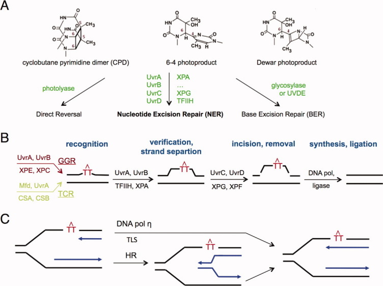
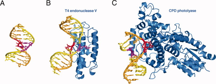
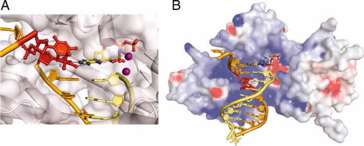
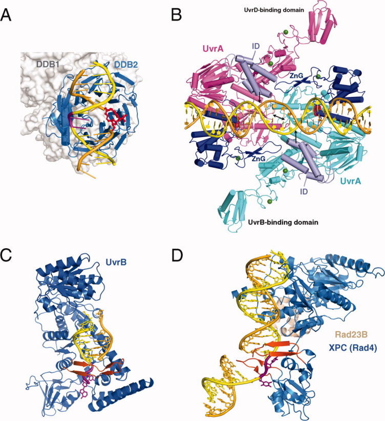
Structural studies of UV-induced lesions and their complexes with repair proteins reveal an intrinsic flexibility of DNA at lesion sites. Reduced DNA rigidity stems primarily from the loss of base stacking, which may manifest as bending, unwinding, base unstacking, or flipping out. The intrinsic flexibility at UV lesions allows efficient initial lesion recognition within a pool of millions to billions of normal DNA base pairs. To bypass the damaged site by translesion synthesis, the specialized DNA polymerase η acts like a molecular "splint" and reinforces B-form DNA by numerous protein-phosphate interactions. Photolyases and glycosylases that specifically repair UV lesions interact directly with UV lesions in bent DNA via surface complementation. UvrA and UvrB, which recognize a variety of lesions in the bacterial nucleotide excision repair pathway, appear to exploit hysteresis exhibited by DNA lesions and conduct an ATP-dependent stress test to distort and separate DNA strands. Similar stress tests are likely conducted in eukaryotic nucleotide excision repair.


Figures

Figure 1
Figure 1
UV lesions and their repair. A: Diagrams of CPD, 6-4 and dewar photoproduct and three different repair pathways. B: An outline of multiple steps in NER. Each step is labeled in bold font and dark blue. UV lesions are represented by red TT with an inverted V above. The NER pathway is divided into global genome (GGR, highlighted in dark red) or transcription coupled repair (TCR, in pale green) based on how lesions are initially recognized. The two branches converge afterwards. Proteins responsible for each step are shown above (bacterial) or below (eukaryotic) each arrow. In the TCR branch RNA polymerase and co-factors are omitted for clarity. C: Bypass of UV lesions during replication is achieved by either translesion synthesis (TLS) or homologous recombination (HR). The template strands are shown in black and daughter strands in blue.
Figure 2
Figure 2
Structures of DNA with UV lesions. A: Cartoon diagram of a naked DNA with a CPD (PDB: 1SM5). The strand with the lesion is shown in orange, and the partner strand in yellow. The CPD is highlighted in red and bases opposite it in magenta. B: The structure of T4 endonuclease V complexed with CPD DNA (PDB: 1VAS). The protein is shown as blue ribbons and the DNA in the same color scheme as in A. C: The structure of photolyase-CPD complex (PDB: 1TEZ).
Figure 3
Figure 3
Human DNA pol η during translesion synthesis. A: A close-up view of the active site with the 3′base of the CPD in the templating position (PDB: 3MR3). The CPD lesion is shown in red; the protein is represented by a silver semitransparent molecular surface. The template strand is colored orange, and the primer yellow. The incoming nucleotide is shown in multicolor and the two Mg2+ essential for the catalysis as purple spheres. Hydrogen bonds between the replicating base pair are represented by gray dotted lines. B: DNA pol η holds the lesion DNA in a straight B-form conformation. DNA is shown in the same color scheme as in A. The molecular surface of Pol η is shown with positive (blue) and negative (red) electrostatic potential.
Figure 4
Figure 4
Proteins involved in NER. A: XPE complexed with 6-4 PP (PDB: 3EI1). The 6-4 PP is highlighted in red and the bases opposite it shown in magenta. DDB2 is a β-propeller protein and shown as blue ribbons. DDB1 binds DDB2 only and is shown in silver molecular surface in the background. B: UvrA-DNA complex (PDB: 3PIH). The bulk of two UvrA subunits are shown in cyan and pink ribbons diagrams except for the ATPase domains that directly contact DNA and contain the C-terminal Zinc-finger domain (dark blue) and the ID domains (pale blue). The UvrB-binding domains are labeled. Pyrophosphates marking the nucleotide-binding site are shown as red spheres, and Zn2+ as green spheres. The unstacked central G:C base pairs are indicated by a black double-headed arrow and the location of fluorescein labeled bases are marked by black arrows. C: UvrB – DNA complex (PDB: 2FDC). The protein is shown as a blue ribbon diagram, and the β hairpin that threads through DNA duplex and clamps down one strand is highlighted in orange-red. The flipped-out bases are highlighted in magenta. They are inserted into a conserved binding pocket. D: The structure of yeast homolog of XPC (Rad4) bound to a UV-damaged DNA. XPC is shown in blue and the partner protein Rad23 in light orange. Two β hairpins (in orange red) “hug” the un-damaged strand, while the damaged bases are flipped out and disordered.
Figure 5
Figure 5
A diagram of lesion recognition and kinetic proofreading by UvrA. UvrA dimer is shown as a symmetric blue open box, and UvrB is shown in green with a β-hairpin. DNA helix (in yellow and orange) binds to UvrA and may be unwound and stretched somewhat, but once released from UvrA (e.g., during UvrA ATP-hydrolysis) it returns to normal B form. But damaged DNA is likely to become bent and unwound under such stress test and undergoes strand separation. UvrB, which is physically attached to UvrA, can then grab the ssDNA and complete lesion recognition.

References

    1. Friedberg EC, Walker GC, Siede W, Wood RD, Schultz RA, Ellenberger T. DNA repair and mutagenesis. 2nd ed. Washington: DC: ASM Press; 2006.
    1. Sancar A. Structure and function of DNA photolyase and cryptochrome blue-light photoreceptors. Chem Rev. 2003;103:2203–2237. - PubMed
    1. Latham KA, Lloyd RS. T4 endonuclease V. Perspectives on catalysis. Ann NY Acad Sci. 1994;726:181–196. discussion 196–187. - PubMed
    1. Goosen N, Moolenaar GF. Repair of UV damage in bacteria. DNA Repair (Amst) 2008;7:353–379. - PubMed
    1. Kaur B, Doetsch PW. Ultraviolet damage endonuclease (Uve1p): a structure and strand-specific DNA endonuclease. Biochemistry. 2000;39:5788–5796. - PubMed

LinkOut - more resources