

Assessment of cortical degeneration in patients with Parkinson's disease by voxel-based morphometry, cortical folding, and cortical thickness
- PMID: 21898679
- PMCID: PMC6870035
- DOI: 10.1002/hbm.21378
Assessment of cortical degeneration in patients with Parkinson's disease by voxel-based morphometry, cortical folding, and cortical thickness
Abstract
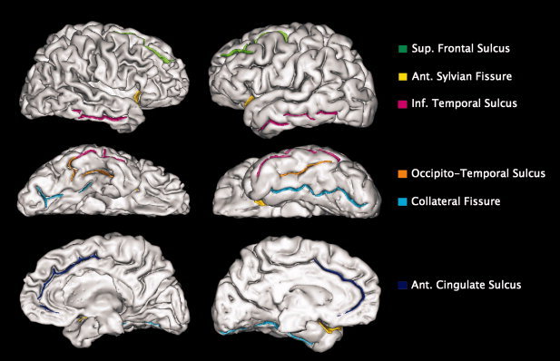
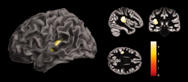
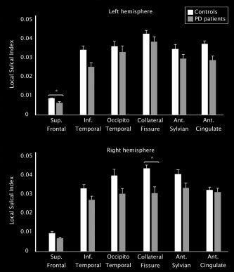
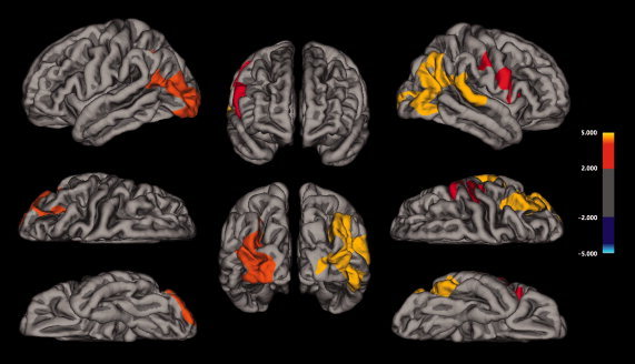
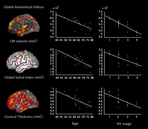
Noninvasive brain imaging methods provide useful information on cerebral involution and degenerative processes. Here we assessed cortical degeneration in 20 nondemented patients with Parkinson's disease (PD) and 20 healthy controls using three quantitative neuroanatomical approaches: voxel-based morphometry (VBM), cortical folding (BrainVisa), and cortical thickness (FreeSurfer). We examined the relationship between global and regional gray matter (GM) volumes, sulcal indices, and thickness measures derived from the previous methods as well as their association with cognitive performance, age, severity of motor symptoms, and disease stage. VBM analyses showed GM volume reductions in the left temporal gyrus in patients compared with controls. Cortical folding measures revealed significant decreases in the left frontal and right collateral sulci in patients. Finally, analysis of cortical thickness showed widespread cortical thinning in right lateral occipital, parietal and left temporal, frontal, and premotor regions. We found that, in patients, all global anatomical measures correlated with age, while GM volume and cortical thickness significantly correlated with disease stage. In controls, a significant association was found between global GM volume and cortical folding with age. Overall these results suggest that the three different methods provide complementary and related information on neurodegenerative changes occurring in PD, however, surface-based measures of cortical folding and especially cortical thickness seem to be more sensitive than VBM to identify regional GM changes associated to PD.
Copyright © 2011 Wiley Periodicals, Inc.
Figures
References
-
- Ashburner J, Friston KJ ( 2000): Voxel‐based morphometry—The methods. Neuroimage 11: 805–821. - PubMed
-
- Ashburner J, Friston KJ ( 2001): Why voxel‐based morphometry should be used. Neuroimage 11: 805–821. - PubMed
-
- Ashburner J, Friston KJ ( 2005): Unified segmentation. Neuroimage 26: 839–851. - PubMed
-
- Ashburner J ( 2007): A fast diffeomorphic image registration algorithm. Neuroimage 38: 95–113. - PubMed
-
- Ashburner J ( 2009): Computational anatomy with the SPM software. Magn Res Imaging 27: 1163–1174. - PubMed
Publication types
MeSH terms
LinkOut - more resources
Full Text Sources
Medical
