

Hydrogen/deuterium exchange and electron-transfer dissociation mass spectrometry determine the interface and dynamics of apolipoprotein E oligomerization
- PMID: 21899263
- PMCID: PMC3202646
- DOI: 10.1021/bi2010027
Hydrogen/deuterium exchange and electron-transfer dissociation mass spectrometry determine the interface and dynamics of apolipoprotein E oligomerization
Abstract
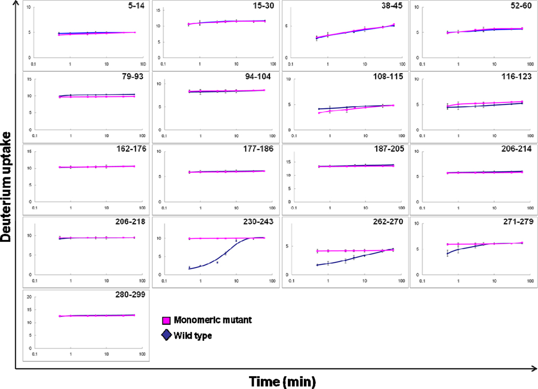
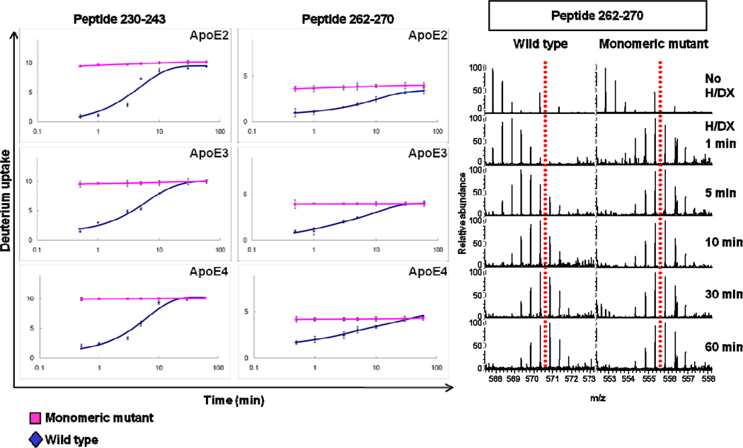
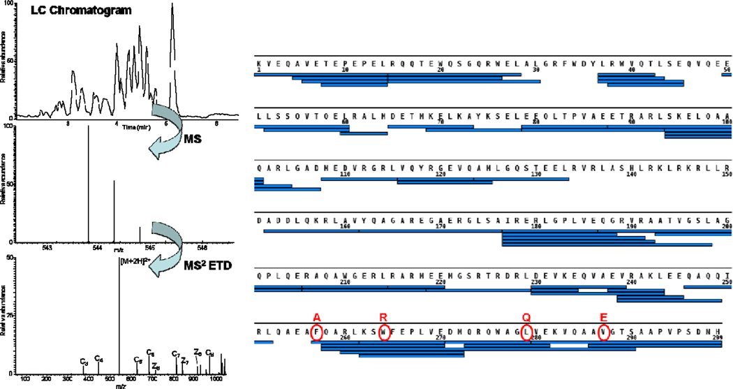
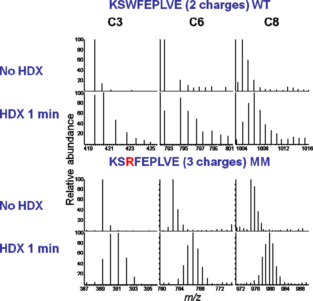
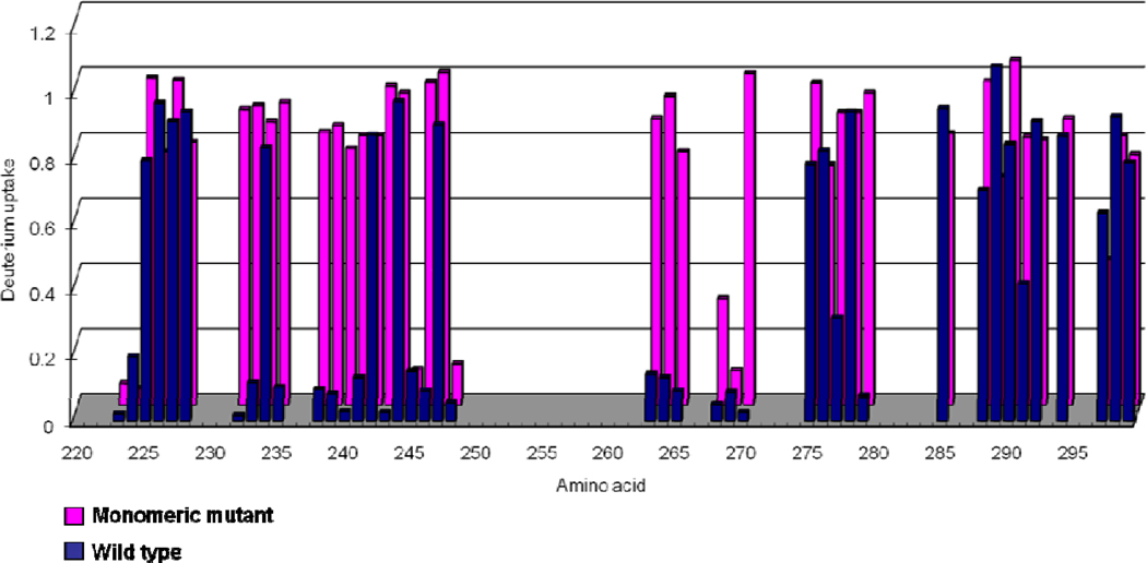
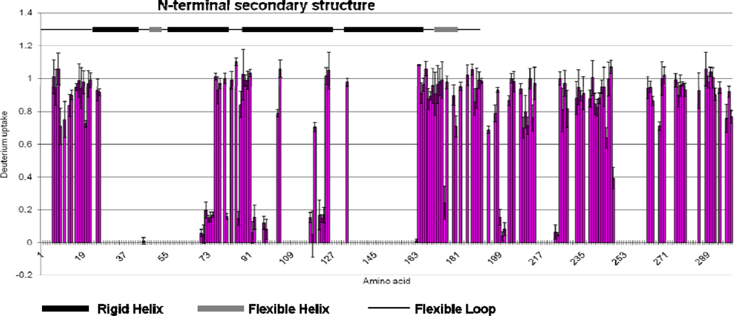
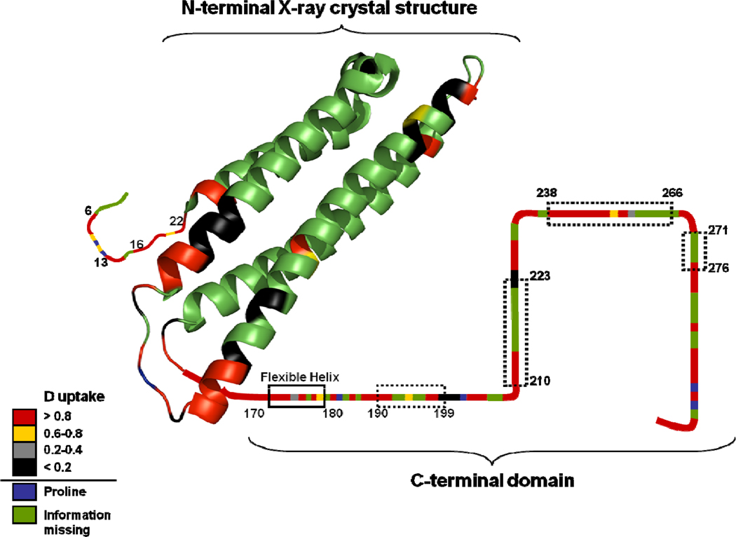
Apolipoprotein E, a 34 kDa protein, plays a key role in triglyceride and cholesterol metabolism. Of the three common isoforms (ApoE2, -3, and -4), only ApoE4 is a risk factor for Alzheimer's disease. All three isoforms of wild-type ApoE self-associate to form oligomers, a process that may have functional consequences. Although the C-terminal domain, residues 216-299, of ApoE is believed to mediate self-association, the specific residues involved in this process are not known. Here we report the use of hydrogen/deuterium exchange (H/DX) coupled with enzymatic digestion to identify those regions in the sequence of full-length apoE involved in oligomerization. For this determination, we compared the results of H/DX of the wild-type proteins and those of monomeric forms obtained by modifying four residues in the C-terminal domain. The three wild-type and mutant isoforms show similar structures based on their similar H/DX kinetics and extents of exchange. Regions of the C-terminus (residues 230-270) of the ApoE isoforms show significant differences of deuterium uptake between oligomeric and monomeric forms, confirming that oligomerization occurs at these regions. To achieve single amino acid resolution, we examined the extents of H/DX by using electron transfer dissociation (ETD) fragmentation of peptides representing selected regions of both the monomeric and the oligomeric forms of ApoE4. From these experiments, we could identify the specific residues involved in ApoE oligomerization. In addition, our results verify that ApoE4 is composed of a compact structure at its N-terminal domain. Regions of C-terminal domain, however, appear to lack defined structure.
Figures
References
-
- Yokoyama S, Kawai Y, Tajima S, Yamamoto A. Behavior of human apolipoprotein E in aqueous solutions and at interfaces. J. Biol. Chem. 1985;260:16375–16382. - PubMed
-
- Hatters DM, Peters-Libeu CA, Weisgraber KH. Apolipoprotein E structure: insights into function. Trends in Biochemical Sciences. 2006;31:445–454. - PubMed
-
- Weisgraber KH, Anfinsen CB, J. T. E. F. M. R., David SE. Advances in Protein Chemistry. Academic Press; 1994. Apolipoprotein E: Structure-Function Relationships; pp. 249–302. - PubMed
-
- Herz J, Beffert U. Apolipoprotein E receptors: linking brain development and alzheimer's disease. Nat. Rev. Neurosci. 2000;1:51–58. - PubMed
-
- Zhang Y, Chen J, Wang J. A complete backbone spectral assignment of lipid-free human apolipoprotein E (apoE) Biomolecular NMR Assignments. 2008;2:207–210. - PubMed
Publication types
MeSH terms
Substances
Grants and funding
LinkOut - more resources
Full Text Sources
Miscellaneous
