. 2011 Sep 7:12:38.
doi: 10.1186/1471-2121-12-38.

The putative Notch ligand HyJagged is a transmembrane protein present in all cell types of adult Hydra and upregulated at the boundary between bud and parent

Affiliations

The putative Notch ligand HyJagged is a transmembrane protein present in all cell types of adult Hydra and upregulated at the boundary between bud and parent

Andrea Prexl et al. BMC Cell Biol. .

Abstract

Background: The Notch signalling pathway is conserved in pre-bilaterian animals. In the Cnidarian Hydra it is involved in interstitial stem cell differentiation and in boundary formation during budding. Experimental evidence suggests that in Hydra Notch is activated by presenilin through proteolytic cleavage at the S3 site as in all animals. However, the endogenous ligand for HvNotch has not been described yet.

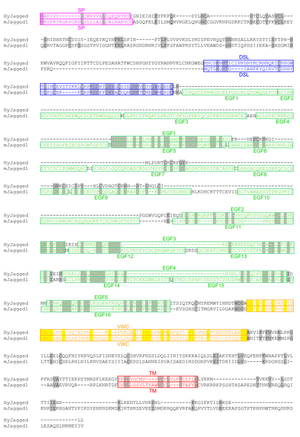
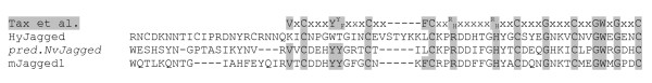
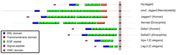
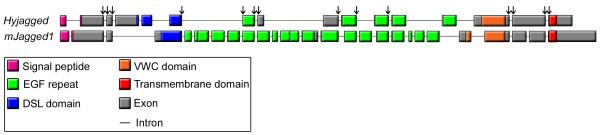
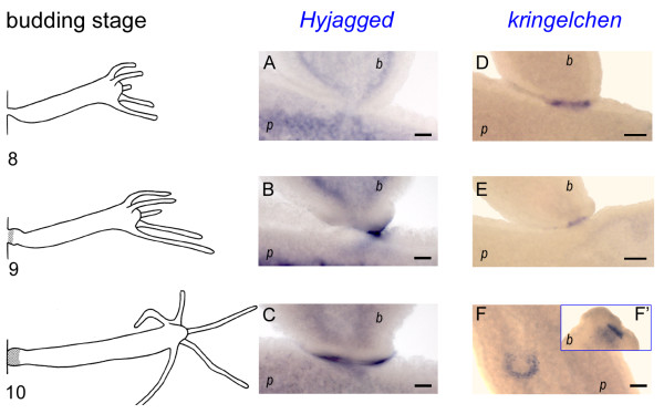
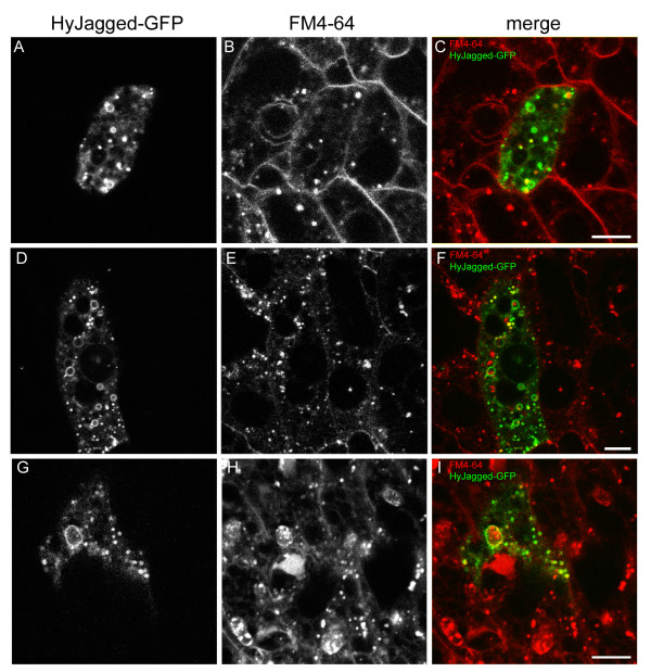
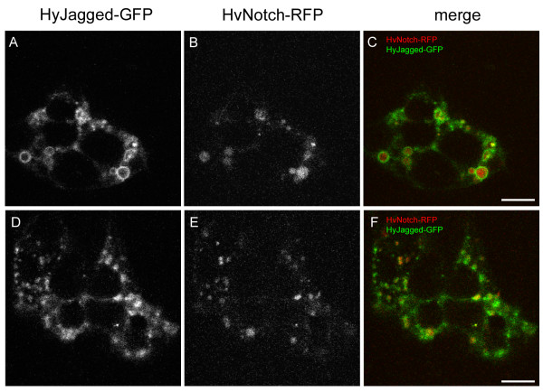
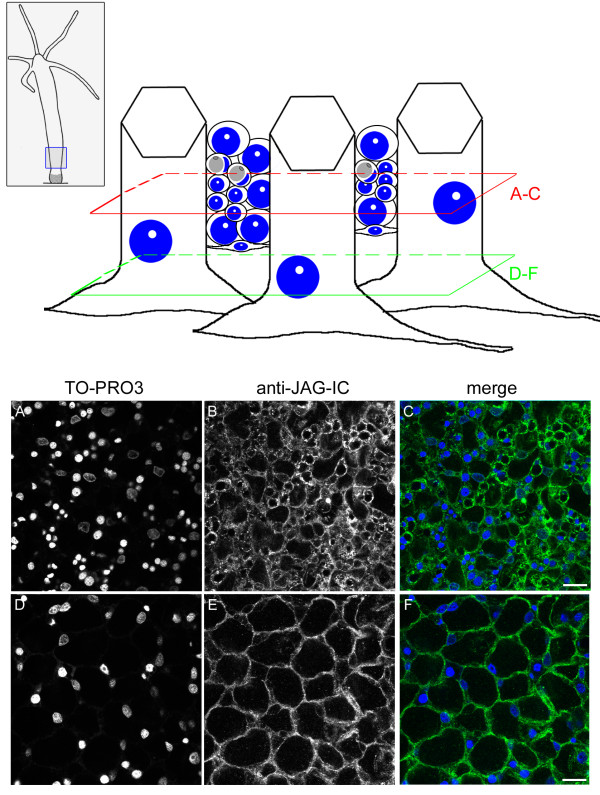
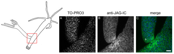
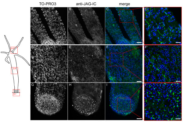
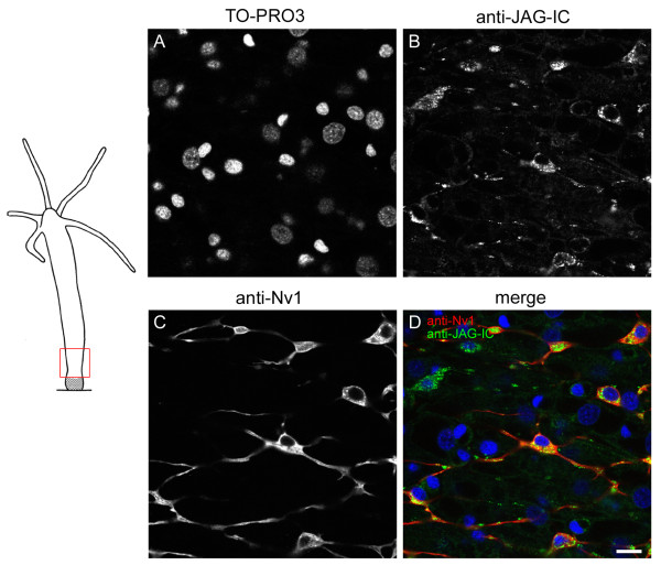
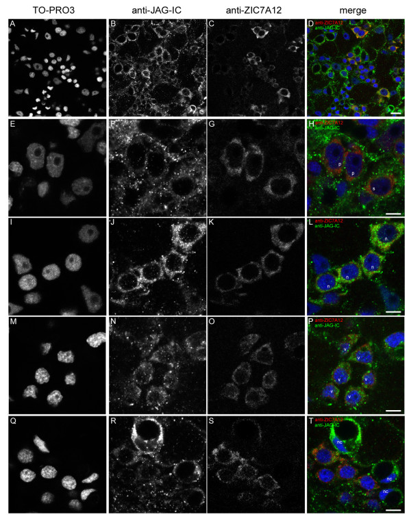
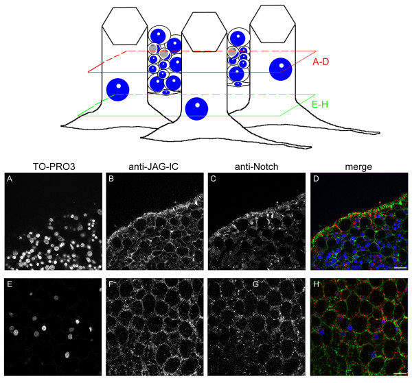
Results: We have cloned a cDNA from Hydra, which encodes a bona-fide Notch ligand with a conserved domain structure similar to that of Jagged-like Notch ligands from other animals. Hyjagged mRNA is undetectable in adult Hydra by in situ hybridisation but is strongly upregulated and easily visible at the border between bud and parent shortly before bud detachment. In contrast, HyJagged protein is found in all cell types of an adult hydra, where it localises to membranes and endosomes. Co-localisation experiments showed that it is present in the same cells as HvNotch, however not always in the same membrane structures.

Conclusions: The putative Notch ligand HyJagged is conserved in Cnidarians. Together with HvNotch it may be involved in the formation of the parent-bud boundary in Hydra. Moreover, protein distribution of both, HvNotch receptor and HyJagged indicate a more widespread function for these two transmembrane proteins in the adult hydra, which may be regulated by additional factors, possibly involving endocytic pathways.


Figures

Figure 1
Figure 1
Clustal X alignment of mJagged1 (mouse) and HyJagged (Hydra). Signal peptide (SP, pink), DSL domain (DSL, blue), EGF repeats (EGF, green), VWC domain (VWC, yellow), transmembrane domain (TM, red). Identical amino acids are highlighted in grey and yellow respectively.
Figure 2
Figure 2
Alignment of DSL domains from predicted NvJagged (Nematostella), mJagged1 (mouse) and HyJagged (Hydra) in comparison with the DSL domain consensus sequence suggested by Tax et al. [13]. Consensus sequence of DSL domains according to [13]. Identical amino acids are marked in grey.
Figure 3
Figure 3
Schematic domain structure of Hydra HyJagged in comparison with Delta and Serrate/Jagged-like proteins from Nematostella, Caenorhabditis, Drosophila and Homo. Signal peptide (pink), DSL domain (blue), EGF repeats (green), VWC domain (orange), transmembrane domain (red).
Figure 4
Figure 4
Schematic representation of the exon-intron structure of Hyjagged (Hydra) in comparison to mJagged1 (mouse). Coding regions for the different protein domains are shown in colour: signal peptide (pink), DSL domain (blue), EGF repeats (green), VWC domain (orange), transmembrane domain (red), non-conserved exon regions (grey). Introns are not to scale (black lines). Conserved exon-intron-boundaries are marked with arrows.
Figure 5
Figure 5
Whole mount in situ hybridisation for Hyjagged and kringelchen during late budding stages. Left hand panel: Budding stages 8, 9 and 10 according to [31]; (A-C) in situ hybridisation for Hyjagged, (D-F') in situ hybridisation for kringelchen from [10]; (F') foot of bud that fell off during staining procedure; (p) parent animal; (b) bud. NBT/BCIP (Roche) was used for staining reactions (blue signals). Scale bars: 20 μm.
Figure 6
Figure 6
Expression of HyJagged-GFP in Hydra. Animals with single HyJagged-GFP expressing cells after transfection with the particle gun were incubated with FM4-64 for 20 min (A-C), 1 h (D-F) and 20 h (G-I); (A, D, G) HyJagged-GFP in a single ectodermal epithelial cell; (B, E, H) FM4-64 in all cells of the animal; (C, F, I) merged images in false colours: HyJagged-GFP (green), FM4-64 (red); single confocal sections; scale bars: 10 μm.
Figure 7
Figure 7
Co-expression of HyJagged-GFP and HvNotch-RFP in Hydra. Two single ectodermal epithelial cells expressing both, HyJagged-GFP and HvNotch-RFP after transfection of animals with the particle gun (cell 1 A-C, cell 2 D-F). (A, D) HyJagged-GFP, (B, E) HvNotch-RFP, (C, F) merged images in false colours: HyJagged-GFP (green), HvNotch-RFP (red); single confocal sections; scale bars: 10 μm.
Figure 8
Figure 8
Immunofluorescence of Hydra whole mounts with the anti-JAG-IC antibody. Upper panel left: schematic representation of an adult hydra, the region imaged in lower panel is indicated, upper panel, right: schematic drawing of cells that were imaged including ectodermal epithelial cells, interstitial cells, nematoblasts, nematocytes and nerve cells; positions of confocal sections are boxed in red (A-C) and green (D-F). (A, D) DNA staining of single confocal sections with TO-PRO3, (B, E) anti-JAG-IC staining, (C, F) merged images in false colours: DNA (blue), anti-JAG-IC staining (green); scale bars: 20 μm.
Figure 9
Figure 9
Staining of HyJagged during budding. Left hand panel: Schematic of an adult hydra with bud at stage 9, region imaged in A-C is indicated; (A) DNA staining of single confocal section with TO-PRO3, (B) anti-JAG-IC staining, (C) merged images of all channels in false colours: DNA (blue), anti-JAG-IC staining (green); scale bar: 50 μm.
Figure 10
Figure 10
Nerve cell staining of HyJagged. Left hand panel: Schematic representation of an adult hydra; regions imaged in A-I are indicated; (A, D, G) DNA staining of stacks of confocal sections with TO-PRO3, (B, E, H) anti-JAG-IC staining, (C, F, I, C', F', I') merged images in false colours: DNA (blue), anti-JAG-IC staining (green); C'F' and I' are magnifications of C, F and I as indicated by red boxes; scale bars: (A-I) 50 μm, (C', F', I') 20 μm.
Figure 11
Figure 11
Co-immunofluorescence staining with anti-JAG-IC- and anti-Nv1-antibodies. Left hand panel: Schematic representation of an adult hydra with imaged region indicated; right hand panel: single confocal sections: (A) DNA staining with TO-PRO3, (B) anti-JAG-IC staining, (C) anti-Nv1 staining, (D) merged images of all channels in false colours: DNA (blue), anti-JAG-IC staining (green), anti-Nv1 staining (red); scale bar: 10 μm.
Figure 12
Figure 12
Co-immunofluorescence staining with anti-JAG-IC- and anti-ZIC7A12-antibodies. Single confocal sections of a hydra from a region of the body column: (A, E, I, M, Q) DNA staining with TO-PRO3, (B, F, J, N, R) anti-JAG-IC staining, (C, G, K, O, S) anti-ZIC7A12 staining, (D, H, L, P, T) merged images of all channels in false colours: DNA (blue), anti-JAG-IC staining (green), anti-ZIC7A12 staining (red); (A-D) overview of the staining pattern in the body column of an adult hydra, (E-H) localization of HyJagged in single (s) and a pair of interstitial cells (p), (I-L) staining of a nest of four nematoblasts (n), (M-P) nest of nematoblasts with vacuoles (v), (Q-T) staining of mature nematocytes (nc); scale bars: (A-D) 10 μm, (E-T) 5 μm.
Figure 13
Figure 13
Co-immunofluorescence staining of Hydra whole mount with anti-JAG-IC- and anti-Notch-antibodies. Upper panel left: schematic representation of an adult hydra, the region imaged in lower panel is indicated; upper panel, right: schematic drawing of cells that were imaged including ectodermal epithelial cells, interstitial cells, nematoblasts, nematocytes and nerve cells; positions of confocal sections are boxed in red (A-D) and green (E-H). (A, E) DNA staining with TO-PRO3, (B, F) anti-JAG-IC staining, (C, G) anti-Notch staining, (D, H) merged images in false colours: DNA (blue), anti-JAG-IC staining (green), anti-Notch staining (red); single confocal sections; scale bars: 20 μm.

References

    1. Hobmayer B, Rentzsch F, Kuhn K, Happel CM, von Laue CC, Snyder P, Rothbächer U, Holstein TW. WNT signalling molecules act in axis formation in the diploblastic metazoan Hydra. Nature. 2000;407:186–189. doi: 10.1038/35025063. - DOI - PubMed
    1. Minobe S, Fei K, Sarras M Jr, Werle M. Identification and characterization of the epithelial polarity receptor "Frizzled" in Hydra vulgaris. Development Genes and Evolution. 2000;210:258–262. doi: 10.1007/s004270050312. - DOI - PubMed
    1. Steele RE. Developmental Signaling in Hydra: What does it take to build a „simple" animal? Developmental Biology. 1996;248:199–219. - PubMed
    1. Hobmayer B, Rentzsch F, Holstein TW. Identification and expression of HySmad1, a member oft he R-Smad family of TGFbeta signal transducers, in the diploblastic metazoan Hydra. Development Genes and Evolution. 2001;211:597–602. doi: 10.1007/s00427-001-0198-8. - DOI - PubMed
    1. Käsbauer T, Towb P, Alexandrova O, David CN, Dall'Armi E, Staudigl A, Stiening B, Böttger A. The Notch signalling pathway in the cnidarian Hydra. Developmental Biology. 2007;303:376–390. doi: 10.1016/j.ydbio.2006.11.022. - DOI - PubMed

Publication types

MeSH terms

LinkOut - more resources