. 2011 Dec;32(35):9197-206.
doi: 10.1016/j.biomaterials.2011.08.059. Epub 2011 Sep 6.

Actin and ERK1/2-CEBPβ signaling mediates phagocytosis-induced innate immune response of osteoprogenitor cells

Affiliations

Actin and ERK1/2-CEBPβ signaling mediates phagocytosis-induced innate immune response of osteoprogenitor cells

Heon Goo Lee et al. Biomaterials. 2011 Dec.

Abstract

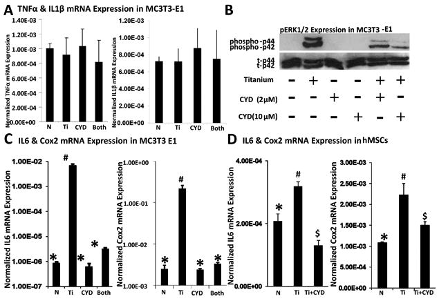
Wear particles at the host bone-implant interface are a major challenge for successful bone implant arthoplasties. Current understanding of aseptic loosening consists of macrophage-mediated inflammatory responses and increasing osteoclastogenesis, which lead to an imbalance between bone formation and resorption. Despite its significant role in bone regeneration and implant osteointegration, the osteoprogenitor response to wear particles has been examined recent years. More specifically, the intracellular mechanism of osteoprogenitor mediated inflammation has not been fully elucidated. In this study, we examined the role of osteoprogenitors and the cellular mechanism by which metal wear particles elicit an inflammatory cascade. Through both in vivo and in vitro experiments, we have demonstrated that osteoprogenitor cells are capable of initiating inflammatory responses by phagocytosing wear particles, which lead to subsequent accumulation of macrophages and osteoclastogenesis, and the ERK_CEBP/β intracellular signaling is a key inflammatory pathway that links phagocytosis of wear particles to inflammatory gene expression in osteoprogenitors. AZD6244 treatment, a potent inhibitor of the ERK pathway, attenuated particle mediated inflammatory osteolysis both in vivo and in vitro. This study advances our understanding of the mechanisms of osteoprogenitor-mediated inflammation, and provides further evidence that the ERK_CEBP/β pathway may be a suitable therapeutic target in the treatment of inflammatory osteolysis.


Conflict of interest statement

Conflict of Interest: All other authors have no conflicts of interest.

Figures

Figure 1
Figure 1. In vivo time course experiments of Ti particle induced osteolysis
Four different groups of mice calvarium were used for histology analyses; Sham, 1 day Ti treated, 3 days Ti treated, and 7 days treated groups. Ti particles (black arrow) were implanted on pericranium (white arrow). All sections of each group were stained with Tri-chrome(top row, A), CD11b (2nd row, B), and TRAP (3rd row, C). (A) Tri-chrome sections were photographed at 10X magnification. (B) CD11b positive cells in pericranium were observed at 40X magnification. (C) TRAP positive osteoclasts (red arrow) at suture line were detected at 40X and (D) quantified at 10X.
Figure 2
Figure 2. In vivo inflammatory responses of pericranium to Ti particle
To investigate the effects of inflammation on osteoclastogenesis, same four groups were used for histology analyses: Sham, 1 day Ti treated, 3 days Ti treated, and 7 days treated groups. All sections of each group were stained with (A) pERK, (B) Cox2, and (C) IL6. The expressions of these inflammation mediators at the pericranium were observed under 40X magnification.
Figure 3
Figure 3. Attachment of Ti particles on cell surface
(A) The schematic of a customized JAVA program for quantification of Titanium (Ti) particle-occupied area on the cell surface is shown. (B) Percentage of Ti-occupied area out of total cell surface is generated based on images from the JAVA program. Different dose of Ti particles were treated to MC3T3-E1 cells with/without 10μm of the Cytochalasin D (CYD) pretreatment. n=20. (C) Actin fluorescent image analysis of particle-occupied area illustrates that Ti particles (Bright View) are accumulated along with actin filaments (Fluorescent image) and 10μm of CYD are able to decrease particle attachment on actin filaments.
Figure 4
Figure 4. Actin remodeling for phagocytosing of Ti particles in osteoprogenitor cell
(A) Actin filaments of selected area (yellow square box) were magnified and projected into X-Y, X-Z, and Y-Z planes by confocal microscopy to visualize actin remodeling. No treatment, Ti, CYD, and Ti+CYD group were compared. Scale bar in non-magnified images is 10μm. Scale bar in magnified images is 1μm. (B) After Ti treatment, vigorous dynamics including actin ring (O) and cup (U) formations were detected in axial, coronal, and sagittal planes. Scale bar represents 1 μm. (C) The quantification analysis of cell associated with Ti particles is shown. Each group has n=300 and * denotes P<0.01.
Figure 5
Figure 5. Ti particle mediated-actin remodeling activates the ERK pathway to induce inflammatory gene expression in MC3T3-E1 and hMSC-derived osteoprogenitor cells
(A) Relative gene expressions of TNFα and IL1β in MC3T3-E1 cells are assessed. No treatment (N), Titanium (Ti), Cytochalasin D (CYD), and Cytochalasin D + Titanium (Both) groups were compared. (B) Immunoblot of MC3T3-E1 cells show the level of phosphorylation of ERK 1/2 (phospho-p44, phosphor-p42) after stimulation of Ti particles with/without CYD. (C&D) Relative gene expressions of IL6 and Cox2 in MC3T3-E1 and hMSC-derived osteoprogenitor cells are assessed. Every gene expression assay is normalized by GAPDH and n=6. Each symbol denotes significant differences (P<0.05).
Figure 6
Figure 6. ERK cascades is a key inflammatory pathway in osteoprogenitor cells
(A) Selective inhibition of pERK1/2 by AZD is assessed through immunoblot. (B) pERK fluorescent image analysis illustrates that (pERK) expression can be induced by titanium particle in MC3T3-E1 and hMSC-derived osteoprogenitor cells. These pERK expressions are also located in both the nucleus and cytoplasm. AZD treatment blocks phosphorylation of ERK expression in both the nucleus and cytoplasm. (C&D) Relative gene expressions of IL6 and Cox2 in MC3T3-E1 and hMSC-derived osteoprogenitor cells are assessed. No treatment (N), Titanium (Ti), AZD (AZD), and AZD + Titanium (Both) groups were compared. Every gene expression assay is normalized by GAPDH and n=6. Each symbol (*, #, $) denotes significant differences (P<0.05).
Figure 7
Figure 7. The ERK pathway mediates phosphorylation of CCAAT/enhancer-binding protein beta (CEBP- β) for inflammatory response to Ti particles
(A) Immunoblot of the cytoplasm and nucleus shows phosphorylated ERK1/2 (phospho-p44, phospho-p42), phosphorylated CEBP-β (phosphor-CEBP-β), GAPDH, and Lamin A/C after Ti particle stimulation with/without AZD or CYD in MC3T3-E1 cells. Effective separations of nuclei and cytoplasm have been confirmed by GAPDH and Lamin A/C detection. (B&C) Prostaglandin E2 (PGE2) and IL6 secretion is measured by ELISA. MC3T3-E1 cells were treated with Ti particles for three different time points (3hr, 6hr and 24hr) in the presence or absence of AZD and CYD pretreatment. Each symbol (*, #, $) denotes significant differences (P<0.05).
Figure 8
Figure 8. The effects of AZD treatment on inflammatory responses to Ti particles in vivo
Four groups were examined to investigate the effects of AZD on inflammation: Sham, 7 day Ti treated, 7 days AZD treated, and 7 days AZD+Ti treated groups. Immunohistochemical staining of pericranium was performed with (A) pERK, (B) Cox2 (C) IL6 and (D) CD11b. Magnification, 40X.
Figure 9
Figure 9. AZD treatment reduces osteoclastogenesis induced by Ti particles in vivo
To investigate the effects of AZD treatment on osteoclastogenesis, quantification of TRAP positive osteoclasts at the suture line was performed at 10X magnification. Representative TRAP positive osteoclasts were photographed from four groups: Sham, 7 day Ti treated, 7 days AZD treated, and 7 days AZD+Ti treated groups. Magnification was set at 40X.
Figure 10
Figure 10
Model for mechanism of inflammatory responses to Ti particles in progenitor cell.

References

    1. Jacobs JJ, Roebuck KA, Archibeck M, Hallab NJ, Glant TT. Osteolysis: basic science. Clin Orthop Relat Res. 2001:71–7. - PubMed
    1. Schmalzried TP, Callaghan JJ. Wear in total hip and knee replacements. J Bone Joint Surg Am. 1999;81:115–36. - PubMed
    1. Harris WH. Wear and periprosthetic osteolysis: the problem. Clin Orthop Relat Res. 2001:66–70. - PubMed
    1. Akisue T, Bauer TW, Farver CF, Mochida Y. The effect of particle wear debris on NFkappaB activation and pro-inflammatory cytokine release in differentiated THP-1 cells. J Biomed Mater Res. 2002;59:507–15. - PubMed
    1. Allen MJ, Myer BJ, Millett PJ, Rushton N. The effects of particulate cobalt, chromium and cobalt-chromium alloy on human osteoblast-like cells in vitro. J Bone Joint Surg Br. 1997;79:475–82. - PubMed

Publication types

MeSH terms