. 2011 Dec;49(3):415-22.
doi: 10.1016/j.molimm.2011.08.005. Epub 2011 Sep 6.

MAPK/AP-1 activation mediates TLR2 agonist-induced SPLUNC1 expression in human lung epithelial cells

Affiliations

MAPK/AP-1 activation mediates TLR2 agonist-induced SPLUNC1 expression in human lung epithelial cells

Jyoti Thaikoottathil et al. Mol Immunol. 2011 Dec.

Abstract

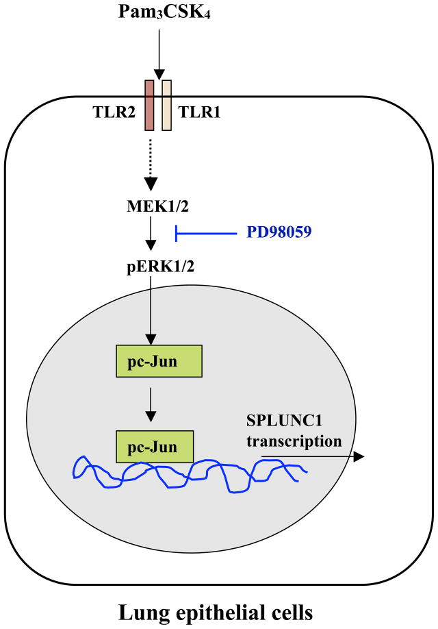
Background: Short Palate Lung and Nasal epithelium Clone 1 (SPLUNC1) is a newly described host defense protein, primarily expressed in large airway epithelial cells. Reduced SPLUNC1 has been reported in allergic and cigarette smoke-exposed airways. We found that Mycoplasma pneumoniae increases SPLUNC1 in airway epithelium in part via activating TLR2-NF-κB pathway. However, the contribution of additional signaling pathways to TLR2-mediated SPLUNC1 expression remains unclear. In the present study, we investigated if TLR2-induced mitogen-activated protein kinase (MAPK)/activator protein-1 (AP-1) signaling regulates SPLUNC1 expression in human lung epithelial cells.

Methods: Human lung epithelial NCI-H292 cells were stimulated with a TLR2 agonist Palmitoyl (3)-Cys-Ser-Lys (4)-OH (Pam(3)CSK(4)). MAPK/AP-1 activation and its role in SPLUNC1 regulation were investigated by Western blot, c-Jun activation assay, chromatin immunoprecipitation (ChIP) and real-time PCR. SPLUNC1 promoter activity was assessed by a luciferase reporter assay.

Results: Pam(3)CSK(4) increased SPLUNC1 expression in NCI-H292 cells in a dose- and time-dependent manner, and enhanced SPLUNC1 promoter activity. Pam(3)CSK(4)-treated cells demonstrated activated MAPK and c-Jun compared to untreated cells. ChIP assay indicated increased c-Jun binding to the SPLUNC1 promoter following Pam(3)CSK(4) stimulation. Inhibition of ERK1/2 significantly reduced Pam(3)CSK(4)-mediated c-Jun activation and SPLUNC1 expression.

Conclusions: Our results for the first time demonstrate that TLR2-mediated MAPK/AP-1 activation up-regulates lung epithelial SPLUNC1 expression at the transcriptional level. Understanding SPLUNC1 gene regulation should provide more specific therapeutic targets to restore deficient SPLUNC1 production in diseased airways.


Figures

FIGURE 1
FIGURE 1. Pam3CSK4 increases SPLUNC1 expression in NCI-H292 cells
NCI-H292 cells were stimulated with Pam3CSK4 after overnight growth in reduced serum medium. SPLUNC1 mRNA and protein expression was determined by real-time PCR and Western blot, respectively. Pam3CSK4 dose-dependently increased SPLUNC1 mRNA at 48 h (A). A time course study revealed a time-dependent increase in SPLUNC1 mRNA by Pam3CSK4 at 100 ng/ml (B). Pam3CSK4 also induced SPLUNC1 protein expression at 48 h as determined by Western blot (C) Data expressed as means ± SEM, n = 6–9.
FIGURE 2
FIGURE 2. Pam3CSK4 increases SPLUNC1 transcriptional activity
NCI-H292 cells were transiently co-transfected with SPLUNC1 promoter reporter construct (−943/+47bp) containing firefly luciferase (F-luc) as reporter gene and renilla luciferase (R-luc) gene for normalizing transfection efficiency. After Pam3CSK4 stimulation for up to 48 h, SPLUNC1 promoter activity was determined by normalizing F-luc to R-luc and expressed as % of Pam3CSK4–treated versus untreated cells (A). To determine the effect of Pam3CSK4 on SPLUNC1 mRNA stability, NCI-H292 cells were stimulated with 100 ng/ml Pam3CSK4 for up to 16 h, and then treated with either 0.1% DMSO or 5 μg/ml actinomycinD (ACD) to inhibit de novo mRNA synthesis. Degradation of SPLUNC1 mRNA was examined for up to 12 h after ACD addition and expressed as % of SPLUNC1 mRNA levels without ACD at indicated time points (B). Data expressed as means ± SEM, n = 3–9.
FIGURE 3
FIGURE 3. Pam3CSK4 activates MAPK/AP-1 in NCI-H292 cells
After overnight growth in reduced serum medium, NCI-H292 cells were stimulated with 100 ng/ml Pam3CSK4 for up to 24 h. Activation of ERK (A) and JNK (B) and total c-Jun expression (C) were determined by Western blot. Upper panel in Figure A, B and C shows densitometry data from 3–6 independent replicates and lower panel is a representative picture of their Western blot. Activation of c-Jun transcription factor, a downstream target of ERK activation, was also determined by using Western blot (D) as well as an ELISA-based TransAM c-Jun activation assay (E). Data expressed as means ± SEM, n = 3–5.
FIGURE 4
FIGURE 4. Pam3CSK4 enhances c-Jun binding to the SPLUNC1 promoter
Several putative AP-1/c-Jun binding motifs were identified in the −2.5kb flanking region of SPLUNC1 gene including SPLUNC1 promoter sequence. Six different primer sets were designed to map 9 putative AP-1/c-Jun binding motifs across −2.5kb flanking region of SPLUNC1 gene (A). After overnight growth in reduced serum medium, NCI-H292 cells were stimulated with Pam3CSK4 (100 ng/ml) for up to 2 h. Binding of c-Jun to SPLUNC1 promoter region (−833/−1036 and −1733/−2004 bases) containing putative Ap-1/c-Jun DNA binding motifs was determined by ChIP assay (B). Upper panel shows a representative picture of agarose gels; lower panel shows densitometric analysis of PCR products visualized on agarose gel electrophoresis. Data expressed as means ± SEM, n = 3.
FIGURE 5
FIGURE 5. Pam3CSK4-mediated ERK1/2 and c-Jun activation up-regulates SPLUNC1 expression
NCI-H292 cells were pre-treated with DMSO (0.1%) or MEK1/2 inhibitor PD98059 (10 μM). An hour later, cells were stimulated with Pam3CSK4 for up to 24 h. ERK1/2 inhibition by PD98059 resulted in decreased ERK1/2 and c-Jun activation as determined by Western blot (A) and ELISA-based TransAM c-Jun activation assay (B). Effect of PD98059 on Pam3CSK4-induced SPLUNC1 mRNA was determined by real-time quantitative PCR (C). Data expressed as means ± SEM, n = 3–5.
FIGURE 6
FIGURE 6. Summary of our research findings
Pam3CSK4-mediated TLR2 activation results in ERK1/2 and c-Jun activation, which enhances c-Jun binding to SPLUNC1 promoter region to up-regulate SPLUNC1 expression in lung epithelial cells.

References

    1. Adiseshaiah P, Kalvakolanu DV, Reddy SP. A JNK-independent signaling pathway regulates TNF alpha-stimulated, c-Jun-driven FRA-1 protooncogene transcription in pulmonary epithelial cells. J Immunol. 2006;177:7193–7202. - PubMed
    1. Bakiri L, Matsuo K, Wisniewska M, Wagner EF, Yaniv M. Promoter specificity and biological activity of tethered AP-1 dimers. Mol Cell Biol. 2002;22:4952–4964. - PMC - PubMed
    1. Ballif BA, Blenis J. Molecular mechanisms mediating mammalian mitogen-activated protein kinase (MAPK) kinase (MEK)-MAPK cell survival signals. Cell Growth Differ. 2001;12:397–408. - PubMed
    1. Bingle CD, Bingle L. Characterisation of the human plunc gene, a gene product with an upper airways and nasopharyngeal restricted expression pattern. Biochim Biophys Acta. 2000;1493:363–367. - PubMed
    1. Chu HW, Gally F, Thaikoottathil J, Janssen-Heininger YM, Wu Q, Zhang G, Reisdorph N, Case S, Minor M, Smith S, Jiang D, Michels N, Simon G, Martin RJ. SPLUNC1 regulation in airway epithelial cells: role of Toll-like receptor 2 signaling. Respir Res. 2010;11:155. - PMC - PubMed

Publication types

MeSH terms

Substances