Does lung ischemia and reperfusion have an impact on coronary flow? A quantitative coronary blood-flow analysis with inflammatory cytokine profile
- PMID: 21900017
- PMCID: PMC3241113
- DOI: 10.1016/j.ejcts.2011.03.053
Does lung ischemia and reperfusion have an impact on coronary flow? A quantitative coronary blood-flow analysis with inflammatory cytokine profile
Abstract
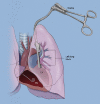
Objective: Ischemia-reperfusion (IR) injury remains a major cause of early morbidity and mortality after lung transplantation with poorly documented extrapulmonary repercussions. To determine the hemodynamic effect due to lung IR injury, we performed a quantitative coronary blood-flow analysis in a swine model of in situ lung ischemia and reperfusion.
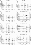
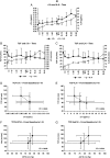
Methods: In 14 healthy pigs, blood flow was measured in the ascending aorta, left anterior descending (LAD), circumflex (Cx), right coronary artery (RCA), right common carotid artery (RCCA), and left internal mammary artery (LIMA), along with left-and right-ventricular pressures (LVP and RVP), aortic pressure (AoP), and pulmonary artery pressure (PAP). Cardiac Troponin (cTn), interleukin 6 and 10 (IL-6 and IL-10), and tumor necrosis factor A (TNF-A) were measured in coronary sinus blood samples. The experimental (IR) group (n=10) underwent 60 min of lung ischemia followed by 60 min of reperfusion by clamping and releasing the left pulmonary hilum. Simultaneous measurements of all parameters were made at baseline and during IR. The control group (n=4) had similar measurements without lung IR.
Results: In the IR group, total coronary flow (TCF=LAD+Cx+RCA blood-flow) decreased precipitously and significantly from baseline (113±41 ml min"1) during IR (p<0.05), with the lowest value observed at 60 min of reperfusion (-37.1%, p<0.003). Baseline cTn (0.08±0.02 ng ml(-1)) increased during IR and peaked at 45 min of reperfusion (+138%, p<0.001). Baseline IL-6 (9.2±2.17 pg ml(-1)) increased during IR and peaked at 60 min of reperfusion (+228%, p<0.0001). Significant LVP drop at 5 min of ischemia (p<0.05) was followed by a slow return to baseline at 45 min of ischemia. A second LVP drop occurred at reperfusion (p<0.05) and persisted. Conversely, RVP increased throughout ischemia (p<0.05) and returned toward baseline during reperfusion. Coronary blood flow and hemodynamic profile remained unchanged in the control group. IL-10 and TNF-A remained below the measurable range for both the groups.
Conclusions: In situ lung IR has a marked negative impact on coronary blood flow, hemodynamics, and inflammatory profile. In addition, to the best of our knowledge, this is the first study where coronary blood flow is directly measured during lung IR, revealing the associated increased cardiac risk.
Figures


Similar articles
-
Remote Ischemic Preconditioning Decreases the Magnitude of Hepatic Ischemia-Reperfusion Injury on a Swine Model of Supraceliac Aortic Cross-Clamping.Ann Vasc Surg. 2018 Apr;48:241-250. doi: 10.1016/j.avsg.2017.08.006. Epub 2017 Sep 6. Ann Vasc Surg. 2018. PMID: 28887256
-
The impact of competitive flow on distal coronary flow and on graft flow during coronary artery bypass surgery.Interact Cardiovasc Thorac Surg. 2011 Jun;12(6):993-7; discussion 997. doi: 10.1510/icvts.2010.255398. Epub 2011 Mar 25. Interact Cardiovasc Thorac Surg. 2011. PMID: 21441253
-
Bronchial microdialysis of cytokines in the epithelial lining fluid in experimental intestinal ischemia and reperfusion before onset of manifest lung injury.Shock. 2010 Nov;34(5):517-24. doi: 10.1097/SHK.0b013e3181dfc430. Shock. 2010. PMID: 20354465
-
Angiotensin-converting enzyme inhibition preserves endothelium-dependent coronary microvascular responses during short-term ischemia-reperfusion.Circulation. 1996 Feb 1;93(3):544-51. doi: 10.1161/01.cir.93.3.544. Circulation. 1996. PMID: 8565174
-
Protection against acute porcine lung ischemia/reperfusion injury by systemic preconditioning via hind limb ischemia.Transpl Int. 2005 Feb;18(2):198-205. doi: 10.1111/j.1432-2277.2004.00005.x. Transpl Int. 2005. PMID: 15691273
Cited by
-
Treatment of acute lung injury by targeting MG53-mediated cell membrane repair.Nat Commun. 2014 Jul 18;5:4387. doi: 10.1038/ncomms5387. Nat Commun. 2014. PMID: 25034454 Free PMC article.
-
A simple method of placing a coronary sinus catheter through the femoral vein in miniature swine.Exp Ther Med. 2017 Apr;13(4):1604-1607. doi: 10.3892/etm.2017.4158. Epub 2017 Feb 22. Exp Ther Med. 2017. PMID: 28413516 Free PMC article.
-
A novel experimental porcine model to assess the impact of differential pulmonary blood flow on ischemia-reperfusion injury after unilateral lung transplantation.Intensive Care Med Exp. 2021 Feb 5;9(1):4. doi: 10.1186/s40635-021-00371-1. Intensive Care Med Exp. 2021. PMID: 33543363 Free PMC article.
References
-
- Jaroszewski D, Hul J, Chu D, Malaisrie C, Riffel A, Gordon H, Wang X, Bakaeen F. Utility of detailed preoperative cardiac testing and incidence of post-thoracotomy myocardial infarction. J Thorac Cardiovasc Surg. 2008;135:648–55. - PubMed
-
- Goldman L, Caldera D, Nussbaum S, Southwick F, Krogstad D, Murray B. Multifactorial index of cardiac risk in noncardiac surgical procedures. N Engl J Med. 1977;297:845–50. - PubMed
-
- Detsky A, Abrams H, Forbath N, Scott J, Hilliard J. Cardiac assessment for patients undergoing noncardiac surgery. A multifactorial clinical risk index. Arch Intern Med. 1986;146:2131–4. - PubMed
-
- Larsen S, Olesen K, Jacobsen E, Nielsen H, Nielsen A, Pietersen A. Prediction of cardiac risk in non-cardiac surgery. Eur Heart J. 1987;8:179–85. - PubMed
Publication types
MeSH terms
Substances
LinkOut - more resources
Full Text Sources
Research Materials

