. 2011 Nov;85(22):11752-69.
doi: 10.1128/JVI.05360-11. Epub 2011 Sep 7.

RelA Ser276 phosphorylation-coupled Lys310 acetylation controls transcriptional elongation of inflammatory cytokines in respiratory syncytial virus infection

Affiliations

RelA Ser276 phosphorylation-coupled Lys310 acetylation controls transcriptional elongation of inflammatory cytokines in respiratory syncytial virus infection

Allan R Brasier et al. J Virol. 2011 Nov.

Abstract

Respiratory syncytial virus (RSV) is a negative-sense single-stranded RNA virus responsible for lower respiratory tract infections (LRTIs) in humans. In experimental models of RSV LRTI, the actions of the nuclear factor κB (NF-κB) transcription factor mediate inflammation and pathology. We have shown that RSV replication induces a mitogen-and-stress-related kinase 1 (MSK-1) pathway that activates NF-κB RelA transcriptional activity by a process involving serine phosphorylation at serine (Ser) residue 276. In this study, we examined the mechanism by which phospho-Ser276 RelA mediates expression of the NF-κB-dependent gene network. RelA-deficient mouse embryonic fibroblasts (MEFs) complemented with the RelA Ser276Ala mutant are deficient in CXCL2/Groβ, KC, and interleukin-6 (IL-6) expression, but NFKBIA/IκBα is preserved. We show that RSV-induced RelA Ser276 phosphorylation is required for acetylation at Lys310, an event required for transcriptional activity and stable association of RelA with the activated positive transcriptional elongation factor (PTEF-b) complex proteins, bromodomain 4 (Brd4), and cyclin-dependent kinase 9 (CDK9). In contrast to gene loading pattern of PTEF-b proteins produced by tumor necrosis factor (TNF) stimulation, RSV induces their initial clearance followed by partial reaccumulation coincident with RelA recruitment. The RSV-induced binding patterns of the CDK9 substrate, phospho-Ser2 RNA polymerase (Pol) II, follows a similar pattern of clearance and downstream gene reaccumulation. The functional role of CDK9 was examined using CDK9 small interfering RNA (siRNA) and CDK inhibitors, where RSV-induced NF-κB-dependent gene expression was significantly inhibited. Finally, although RSV induces a transition from short transcripts to fully spliced mRNA in wild-type RelA (RelA WT)-expressing cells, this transition is not seen in cells expressing RelA Ser276Ala. We conclude that RelA Ser276 phosphorylation mediates RelA acetylation, Brd4/CDK9 association, and activation of downstream inflammatory genes by transcriptional elongation in RSV infection.


Figures
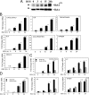
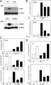
FIG. 1.
FIG. 1.
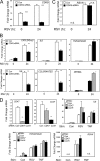
Inducible RelA Ser276 phosphorylation mediates expression of a network of cytokine genes. (A) A549 cells were infected with sucrose-purified RSV (MOI, 1.0) for 0 to 24 h as indicated (top) and lysed, and equal amounts of whole-cell extract were resolved by 8% SDS-PAGE and transferred to a polyvinylidene difluoride (PVDF) membrane. The membrane was incubated with either anti-phospho-Ser276 RelA Ab (RelAp276) (upper panel) or anti-RelA Ab, recognizing total RelA (RelA) (lower panel). Migration of molecular mass markers (in kDa) is shown on the left. (B) A549 cells were RSV infected, and total RNA was isolated for gene expression analysis by Q-RT-PCR. Shown are fold changes in CXCL2/Groβ, IL-8, TNFAIP3/A20, IL-6, CCL5/RANTES, and NFKBIA/IκBα mRNAs normalized to GAPDH after 0, 6, 15, and 24 h of RSV infection. *, P < 0.05; **, P < 0.01. (C) RelA-deficient MEFs stably expressing EGFP-RelA WT or EGFP-RelA Ser276Ala mutant proteins were RSV infected (MOI, 1.0). Shown are fold changes in mCXCL2/Groβ, KC, IL-6, and NFKBIA. (D) Cellular RNA from RSV-infected cells was extracted at indicated times and assayed for expression of RSV N. n.d., not detected; n.s., not significant.
FIG. 2.
FIG. 2.
RSV-induced RelA acetylation at Lys310 is dependent on phosphorylation at Ser276. (A) A549 cells were infected with RSV as indicated, and whole-cell extracts were prepared and immunoprecipitated with anti-RelA antibodies. RelA acetylation at Lys310 was determined by Western blotting using anti-Ac-Lys310 RelA-specific Ab. (B) A549 cells stably expressing EGFP-RelA (F-RelA) were infected with RSV (MOI, 1.0) for the indicated times. TNF stimulation (0 to 60 min) served as a positive control. EGFP-RelA was affinity isolated, and acetylation of RelA at Lys310 was detected in Western blotting with anti-Ac-Lys310 RelA-specific Ab (upper panel). Blots were probed with anti-RelA Ab detecting total RelA as loading control (bottom panel). Migration of molecular mass markers (in kDa) is shown at left. (C) RelA−/− MEFs stably expressing EGFP-RelA WT or the RelA Ser276Ala mutant were infected with RSV and affinity purified. Shown is a Western blot of the immunoprecipitates probed with anti-Ac-Lys310 RelA Ab (top panel) or anti-RelA antibody (bottom panel).
FIG. 3.
FIG. 3.
RelA acetylation on Lys310 is required for inducible transactivation of NF-κB-dependent target genes. (A) Western immunoblot of RelA mutants expressed in RelA−/− MEFs. EGFP-RelA WT-, EGFP-RelA Ser276Ala-, and EGFP-RelA Lys310Arg-expressing cells were lysed and RelA abundance quantified by Western immunoblotting using anti-RelA Ab. The blot was reprobed with anti-β-actin Ab as loading control (bottom panel). (B) Confocal images of control or dsRNA-stimulated MEFs expressing RelA WT (top) or RelA Lys310Arg (bottom). For each image, DAPI staining indicates the location of nuclei. (C) Quantification of nuclear translocation. The nuclear/cytoplasmic ratio of EGFP-RelA was extracted by automated image analysis (CellProfiler) (see Materials and Methods) and plotted as a histogram. The vertical arrow indicates the population median value. Note the shift to the right of the median nuclear/cytoplasmic ratio in the dsRNA-stimulated cells. (D) XChIP was performed on RelA−/− MEFs stably transfected with EGFP-RelA Lys310Arg. Shown is the fold change in mCXCL2/Groβ promoter occupancy in response to RSV infection. Immunoprecipitation with IgG was the negative control. (E) RelA−/− MEFs stably expressing EGFP-RelA WT or EGFP-RelA Ser276Ala mutant proteins were RSV infected (MOI, 1.0). Shown are fold changes in mCXCL2/Groβ, KC, IL-6, and NFKBIA. (F) RelA−/− MEFs expressing RelA WT or RelA Lys310Arg were RSV infected (MOI, 1.0). Cellular RNA from RSV-infected cells was extracted at the indicated times and assayed for expression of RSV N. n.d., not detected; n.s., not significant. (G) One hundred micrograms of A549 cells stably expressing nothing (−), EGFP-RelA WT, EGFP-RelA S276A (276A), or EGFP-RelA Lys310Arg (310R) F-RelA was fractionated by SDS-PAGE and subjected to Western blotting. Top, Western immunoblot probed with anti-RelA Ab. The ∼100-kDa EGFP-RelA and endogenous 65-kDa RelA are indicated. Bottom, the blot was reprobed with anti-β-actin Ab as a loading control. (H) A549 cells stably expressing EGFP-RelA WT, EGFP-RelA Ser276Ala, or EGFP-RelA Lys310Arg proteins were RSV infected (MOI, 1.0, 24 h). Shown are fold change in hCXLC2/Groβ, IL-8, and IL-6 mRNAs as determined by Q-RT-PCR. Experiments were repeated twice.
FIG. 4.
FIG. 4.
RSV-inducible Brd4 association with RelA is dependent on RelA phosphorylation on Ser276. (A) RelA−/− MEF cells expressing either EGFP-RelA WT or EGFP-RelA Ser276Ala were infected or not with RSV for 15 h. Equal amount of extracts were affinity purified, and immunoprecipitates were assayed for Brd4 binding by Western blotting with probing with anti-Brd4-Ab (top panel) or anti-CDK9 Ab (bottom panel). The membrane was stripped and reprobed with anti-RelA Ab as a loading control. (B) A549 cells were stimulated with TNF for the indicated times and assayed by XChIP with IgG, anti-RelA (top), or anti-Brd4 Ab (bottom panel). Shown is Q-gPCR for fold enrichment of the CXCL2/Groβ promoter. (C) XChIP assay using anti-RelA (top), Brd4 (middle), and acetyl-Lys5 histone H4 (H4AcK5) (bottom) Abs to the CXCL2/Groβ promoter for various times after RSV infection (in hours). (D) RSV-infected A549 cells were subjected to XChIP for Brd4 and Ac-Lys5 H4 recruitment to the IL-8 promoter. Note the rapid appearance of Ac-Lys5 H4, which precedes the reaccumulation of Brd4 binding.
FIG. 5.
FIG. 5.
Inducible CDK9 and phospho-Ser2 CTD Pol II loading on NF-κB-dependent genes. (A) Diagram of the exon (E)-intron (I) topology of the human CXCL2/Groβ gene. The locations of the 5′-, intron 3 (I3)-, and 3′-directed primers for Q-gPCR assay are shown. (B) A549 cells were stimulated with TNF for the indicated times. Chromatin was isolated and immunoprecipitated with anti-CDK9 Ab (top) or anti-phospho-Ser2 CTD RNA Pol II Ab (bottom). Shown is Q-gPCR using region-specific XChIP primers as indicated at the top. Note that the CDK9 and phospho-Ser2 CTD Pol II are induced on the 5′ region of the gene and later appear in intron 3 and the 3′UTR. (C) A549 cells were RSV infected for the indicated times (in hours) and subjected to region-specific XChIP for CDK9 (top) and phospho-Ser2 RNA Pol II (bottom) using the indicated Q-gPCR primers. Note the similar patterns of CDK9 and phospho-Ser2 RNA Pol II clearance and reaccumulation on the 5′ region as seen for Brd4. (D) A549 cells were stimulated with 300-bp dsRNA for the indicated times (in hours). XChIP assays were performed using anti-RelA Ab for the proximal promoter of the CXL2/Groβ gene. (E) Region-specific CDK9 and phospho-Ser2 CTD RNA Pol II XChIP was performed as for panel C. Note the rapid clearance of CDK9 and reaccumulation with the loading of phospho-Ser2 RNA Pol II on intron 3 and the 3′UTR of the gene. (F) TNF-stimulated A549 cells were subjected to XChIP assays using anti-CDK7 (top) and cyclin H (bottom) Abs. Shown is proximal CXCL-2/Groβ promoter binding as determined by Q-gPCR for the indicated times. (G) RSV-infected A549 cells were subjected to XChIP for CDK7 and cyclin H and assayed for proximal CXCL-2/Groβ promoter enrichment.
FIG. 6.
FIG. 6.
RSV-inducible cytokine expression is CDK9 dependent. (A) Control and CDK9 siRNAs were transfected into A549 cells, and 48 h later, cells were infected with or without RSV (MOI, 1.0) for another 24 h. The total RNAs were collected for gene expression analysis using Q-RT-PCR. Shown is the fold change in CDK9 gene expression. (B) Effect of CDK9 knockdown on RSV-inducible NF-κB-dependent gene expression. RNA prepared from the experiment for panel A was analyzed for expression of CXL2/Groβ, IL-8, IL-6, NFKBIA, TNFAIP3/A20, and CCL5/RANTES by Q-RT-PCR. Shown are fold changes in mRNA expression normalized to GAPDH expression. *, P < 0.05; **, P < 0.01. (C) Total RNA from the same experiment was analyzed for RSV replication. Shown is Q-RT-PCR for RSV N expression. n.d., not detected; n.s., not significant. (D) Effect of CDK7 and cyclin H knockdown. A549 cells transfected with control siRNA or siRNAs specific to CDK7 and cyclin H were stimulated with TNF or infected with RSV. Shown is mRNA expression of IL-6, TNFAIP3/A20, and RSV N as determined by Q-RT-PCR. **, P < 0.01 compared to control values; n.d., not detected.
FIG. 7.
FIG. 7.
CDK inhibitors block RSV-inducible gene expression. (A) A549 cells were pretreated with vehicle or FP (500 nM) for 1 h prior to different periods of RSV infection. Total RNAs were processed for analysis of gene expression by Q-RT-PCR. Shown are fold changes in CXCL2/Groβ, IL-8, TNFAIP3/A20, IL-6, CCL5/RANTES, and NFKBIA mRNAs normalized to GAPDH after 0, 6, 15, and 24 h of RSV infection. *, P < 0.05; **, P < 0.01. (B) A549 cells were pretreated with vehicle or FP (500 nM) for 1 h prior to RSV infection. The conditioned medium was collected for cytokine determination by multiplex ELISA. Shown are the changes in IL-8, MCP1, IP-10, IL-6, and CCL5/RANTES levels.
FIG. 8.
FIG. 8.
A CDK9 inhibitor blocks RSV-inducible phospho-Ser2 CTD Pol II recruitment and cytokine expression. (A) A549 cells treated with increasing concentrations of CAN508 (100 μM and 200 μM) were RSV infected. At 24 h, total RNA was extracted and subjected to Q-RT-PCR for expression of CXCL2/Groβ, IL-8, TNFAIP3/A20, IL-6, CCL5/RANTES, and NFKBIA mRNAs. Shown are fold changes in mRNA expression normalized to GAPDH. **, P < 0.01. (B) RNA samples from the experiment of panel A were analyzed for changes in RSV N transcription. (C) Effect of CAN508 on RSV-inducible phospho-Ser2 RNA Pol II recruitment. A549 cells were treated in the absence or presence of CAN508 (200 μM) and infected or not with RSV (24 h; MOI, 1.0). Chromatin was cross-linked and immunoprecipitated with the indicated Ab. Shown is the fold change in CXLC2/Groβ using the 5′ primer set. *, P < 0.05.
FIG. 9.
FIG. 9.
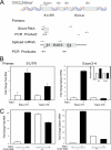
RSV-inducible transcriptional elongation is phospho-Ser276A RelA dependent. (A) Schematic diagram of mCXCL2/Groβ RNA transcripts. The annealing regions of two primer pairs are shown. The 5′UTR primer pair amplifies the 5′UTR of mCXCL2/Groβ RNA, producing a product for abortive and spliced mRNAs. In contrast, the exon 3-4 primer pair produces a real-time PCR signal only in the presence of the spliced mRNA. (B) RelA-deficient MEFs stably expressing EGFP-RelA WT or the RelA Ser276Ala mutant were treated in the absence or presence or RSV. Total RNA was extracted and subjected to Q-RT-PCR analysis using the 5′UTR and exon 3-4 primer pairs. Shown is the fold change for each. Inset, elongation index, determined by the ratio of absolute concentration of total transcripts minus spliced transcripts to the total transcripts. Note that the elongation ratio increases in cells expressing RelA WT but does not change in cells expressing the RelA Ser276Ala mutation. (C) RelA-deficient MEFs stably expressing EGFP-RelA WT were exposed to RSV in the absence or presence of CAN508. Shown is the fold change in mCXCL2/Groβ as determined by Q-RT-PCR analysis using the 5′UTR and exon 3-4 primer pairs. Note that the expression of mCXCL2/Groβ spliced transcript is completely inhibited.
FIG. 10.
FIG. 10.
Effect of RSV-induced phospho Ser276 RelA formation on cytokine expression. Shown is a schematic model of the effect of phospho-Ser276 RelA formation on inducible transcriptional elongation. RSV induces MSK-1 through an ROS-dependent mechanism. MSK1 is required for RelA Ser276 phosphorylation, a posttranslational modification required for binding of Brd4 and CDK9. Brd4 and CDK9 binding to target gene promoters is cleared, followed by a RelA-dependent recruitment of Brd4 and CDK9 to promoters coincident with target gene expression. CDK9 phosphorylates Ser2 in the heptad repeat of CTD of RNA Pol II, resulting in transcriptional elongation and target gene expression.

Similar articles

Cited by

References

    1. Aherne W. T., Bird T., Court S. D. B., Gardner P. S., McQuillin J. 1970. Pathological changes in virus infections of the lower respiratory tract in children. J. Clin. Pathol. 23: 7–18 - PMC - PubMed
    1. Akagi T., Sasai K., Hanafusa H. 2003. Refractory nature of normal human diploid fibroblasts with respect to oncogene-mediated transformation. Proc. Natl. Acad. Sci. U. S. A. 100: 13567–13572 - PMC - PubMed
    1. Anrather J., Racchumi G., Iadecola C. 2005. cis-acting element-specific transcriptional activity of differentially phosphorylated nuclear factor-κB. J. Biol. Chem. 280: 244–252 - PubMed
    1. Barnes P. J., Karin M. 1997. Nuclear factor-kappaB: a pivotal transcription factor in chronic inflammatory diseases. N. Engl. J. Med. 336: 1066–1071 - PubMed
    1. Brasier A. R. 2008. Expanding role of cyclin dependent kinases in cytokine inducible gene expression. Cell Cycle 7: 1–6 - PMC - PubMed

Publication types

LinkOut - more resources