. 2011 Nov 3;118(18):5031-9.
doi: 10.1182/blood-2011-07-365247. Epub 2011 Sep 6.

Inhibition of IL-32 activation by α-1 antitrypsin suppresses alloreactivity and increases survival in an allogeneic murine marrow transplantation model

Affiliations

Inhibition of IL-32 activation by α-1 antitrypsin suppresses alloreactivity and increases survival in an allogeneic murine marrow transplantation model

A Mario Marcondes et al. Blood. .

Abstract

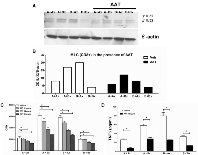
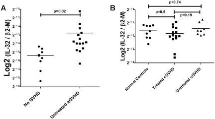
Interleukin (IL)-32 was originally identified in natural killer cells and IL-2-activated human T lymphocytes. As T cells are activated in allogeneic transplantation, we determined the role of IL-32 in human mixed lymphocyte cultures (MLCs) and GVHD. In allogeneic MLCs, IL-32 increased two-fold in responding T cells, accompanied by five-fold increases of TNFα, IL-6, and IL-8. After allogeneic hematopoietic cell transplantation, IL-32 mRNA levels in blood leukocytes were statistically significantly higher in patients with acute GVHD (n = 10) than in serial samples from patients who did not develop acute GVHD (n = 5; P = .02). No significant changes in IL-32 levels were present in patients with treated (n = 14) or untreated (n = 8) chronic GVHD, compared with healthy controls (n = 8; P = .5, and P = .74, respectively). As IL-32 is activated by proteinase-3 (PR3), we determined the effect of the serine protease inhibitor α-1 antitrypsin (AAT) on IL-32 levels and showed suppression of IL-32 and T-lymphocyte proliferation in MLCs. In an MHC-minor antigen disparate murine transplant model, preconditioning and postconditioning treatment with AAT resulted in attenuation or prevention of GVHD and superior survival compared with albumin-treated controls (80% vs 44%; P = .04). These findings suggest that AAT modulates immune and inflammatory functions and may represent a novel approach to prevent or treat GVHD.


Figures

Figure 1
Figure 1
Mixed leukocyte culture (MLC) and IL-32 expression. (A) PBMCs were cultured for 7 days, and Western blots were generated from either unsorted or sorted CD8+ responder cells; 1 blot is representative of 3 similar experiments. (B) IL-32 mRNA levels in allogeneic MLCs. Error bars represent mean ± SEM of 3 similar experiments. Cytokines from MLC supernatants were determined by ELISA, including TNF-α (C), IL-6 (D), and IL-8 (E). Solid columns represent results in allogeneic cultures, open columns represent results in autologous controls. The results are displayed as ± SEM from 3 similar experiments (*P < .01; Student t test).
Figure 2
Figure 2
Effect of IL-32 specific siRNA and AAT on expression of inflammatory mediators. (A) Change in cytokine expression in PBMCs transfected with IL-32–specific or scrambled siRNA (control). Cytokine expression was assayed using profiler cytokine array (R&D Systems). Cytokine concentrations from siRNA transfected PBMC supernatants are expressed as percent change in comparison to control supernatants. Shown are changes (mean ± SEM) in 29 cytokines. The horizontal line indicates a decrease of 25% in comparison to controls transfected with scrambled sequence. Levels were determined after 72 hours of culture. The membrane contained probes for C5a, ICAM-1, IL-4, IL-13, IL-32α, MIP-1β, CD40 ligand, IFN-γ, IL-5, IL-16, IP-10, RANTES, G-CSF, IL-1α, IL-6, IL-17, I-TAC, SDF-1, GM-CSF, IL-1β, IL-8, IL-17E, MCP-1, Serpin-E1, GROα, IL-1ra, IL-10, IL-23, MIF, TNFα, I-309, IL-2, IL-12p70, IL-27 MIP-1α, and TREM-1. (B) Western blot of protein extract of the human stroma cell line HS5 exposed to vehicle only (veh) or various concentrations of AAT (in serum-free medium). Shown are levels of IL-32 β and γ isoforms at concentrations of ATT between 0.1 and 1 mg/mL. This blot is representative of 3 similar experiments.
Figure 3
Figure 3
Inhibition of proliferation and TNFα secretion in MLCs by AAT. (A) Western blot of IL-32β and γ levels in CD8+ cells from 7-day MLCs under control conditions and in the presence of AAT (0.3 mg/mL). IL-32 β and γ isoforms in the presence of AAT. The Western blot is representative of 3 similar experiments. (B) Expression changes in IL-32 protein levels in allogeneic MLCs and autologous controls as determined by densitometry (OD) of the same biologic experiment. Open columns reflect results in the absence of AAT; solid columns in the presence of AAT. (C) Proliferation in MLCs (as measured by 3H thymidine uptake; CPM, mean ± SEM). (D) TNF-α ELISA. Secretion of TNF α in the presence and absence of AAT (*P < .05; Student t test).
Figure 4
Figure 4
IL32 mRNA levels in WBCs and PBMCs from patients with or without clinical GVHD after hematopoietic cell transplantation. (A) Acute GVHD. IL-32 expression was determined with probe Hs00170403_m1 covering all isoforms (see “Analysis of human and murine cytokines by real-time PCR”) and validated using 4 different taqman probes for IL-32 (data not shown). Expression of IL-32 in WBCs from patients with acute GVHD (10 patients, 15 samples) was twice as high as in patients who did not develop clinical signs of GVHD (5 patients, 8 samples; P < .02). (B) Chronic GVHD. Steady-state levels of IL-32 mRNA in PBMCs from patients with chronic GVHD. No significant changes in IL-32 expression were seen in PBMCs of patients with chronic GVHD either untreated (n = 8, P = .74) or treated (n = 14, P = .5) compared with healthy controls. Similarly, there was no difference between treated and untreated patients with chronic GVHD (P = .19; Student t test for comparison of continuous variables between 2 groups; 1-way ANOVA for 3 or more groups).
Figure 5
Figure 5
Effect of AAT on GVHD severity and mortality. (A) AAT treatment scheme. (B) Survival. Survival of AAT-treated mice versus albumin-treated controls (n = 15 each group, P = .04). (C) Severity of GVHD. GVHD was scored based on percentage of weight loss, skin integrity, posture, mobility, and fur texture. Clinical signs were graded on a scale of 0 to 2, where 0 was absent, 1 was moderate, 2 was severe, and the individual scores were added. Shown are GVHD clinical scores for 30 days after transplantation (mean ± SEM per time point; D) Change in body weight of transplanted mice over time after transplantation (mean ± SEM; n = 15). (E) Donor chimerism. Proportion of donor cells among PBMCs in AAT-treated (n = 6) versus albumin-treated (n = 5) mice at day 45 (P = .25).
Figure 6
Figure 6
Skin, stomach, and duodenal histology in AAT-treated and albumin-treated C57/BL6J mice transplanted from C3H.SW-H2b/SnJ donors. Mice were sacrificed on day 21 after transplantation (3 mice per group). Tissues were placed in 10% formalin, embedded in paraffin, sectioned, stained with H&E, and analyzed. Slides were imaged on an Axio Observer inverted microscope (Carl Zeiss MicroImaging) and captured with an Axiocam MRm camera (Carl Zeiss MicroImaging). (A) Skin section of albumin-treated mouse showing edema with interstitial inflammation (10×). (B) Skin section of albumin-treated mouse showing interstitial fibrosis and inflammation invading and damaging hair follicles, characteristic of GVHD (10× lens). (C-D) Skin of 2 AAT-treated mice showing normal histology (10× and 4×, respectively). (E) Stomach of albumin-treated mouse showing interstitial gastritis and evidence of GVHD. (F) Stomach of albumin-treated mouse showing interstitial gastritis and evidence of GVHD with tissue edema and lymphocytic infiltration. (G) Forestomach of AAT-treated mouse showing minimal evidence of GVHD (10×). (H) Stomach of AAT-treated mouse showing normal histology. (I-J) Glandular forestomach/duodenum of albumin-treated mouse showing interstitial inflammation, crypt loss, and apoptotic bodies with 1 small crypt abscess, typical for GVHD (4× and 10×). (K-L) Duodenum of AAT-treated mouse with minimal focal evidence of GVHD (4× and 10×).
Figure 7
Figure 7
Effect of AAT on cytokine RNA and protein expression in PBMCs and plasma after transplantation. (A) IL-1Ra, IL-1β, TNF-α, and PR3 RNA levels, determined by RT-PCR, in PBMCs. Levels in AAT-treated mice (n = 6) are expressed relative to levels in albumin treated controls; mean ± SEM (n = 6; log2). (B-D) Mean ± SEM cytokine plasma levels at 3, 7, and 10 days after transplantation. Shown is a panel selected from a mouse array of 40 cytokines, showing significant changes. Changes in cytokine concentration are expressed as percent change compared with albumin control. The horizontal dotted line indicates an increase/decrease of 25%.

References

    1. Antin JH, Ferrara JLM. Cytokine dysregulation and acute graft-versus-host disease. Blood. 1992;80(12):2964–2968. - PubMed
    1. Stirewalt DL, Mhyre AJ, Marcondes M, et al. Tumour necrosis factor-induced gene expression in human marrow stroma: clues to the pathophysiology of MDS? Br J Haematol. 2008;140(4):444–453. - PubMed
    1. Marcondes AM, Mhyre AJ, Stirewalt DL, Kim S-H, Dinarello CA, Deeg HJ. Dysregulation of IL-32 in myelodysplastic syndrome and chronic myelomonocytic leukemia modulates apoptosis and impairs NK function. Proc Natl Acad Sci U S A. 2008;105(8):2865–2870. - PMC - PubMed
    1. Hong J, Bae S, Kang Y, et al. Suppressing IL-32 in monocytes impairs the induction of the proinflammatory cytokines TNFalpha and IL-1beta. Cytokine. 2010;49(2):171–176. - PubMed
    1. Mun SH, Kim JW, Nah SS, et al. Tumor necrosis factor alpha-induced interleukin-32 is positively regulated via the Syk/protein kinase Cdelta/JNK pathway in rheumatoid synovial fibroblasts. Arthritis Rheum. 2009;60(3):678–685. - PubMed

Publication types

MeSH terms