. 2011 Oct 28;286(43):37615-24.
doi: 10.1074/jbc.M111.268029. Epub 2011 Sep 7.

The RhoG/ELMO1/Dock180 signaling module is required for spine morphogenesis in hippocampal neurons

Affiliations

The RhoG/ELMO1/Dock180 signaling module is required for spine morphogenesis in hippocampal neurons

Jeong-Yoon Kim et al. J Biol Chem. .

Abstract

Dendritic spines are actin-rich structures, the formation and plasticity of which are regulated by the Rho GTPases in response to synaptic input. Although several guanine nucleotide exchange factors (GEFs) have been implicated in spine development and plasticity in hippocampal neurons, it is not known how many different Rho GEFs contribute to spine morphogenesis or how they coordinate the initiation, establishment, and maintenance of spines. In this study, we screened 70 rat Rho GEFs in cultured hippocampal neurons by RNA interference and identified a number of candidates that affected spine morphogenesis. Of these, Dock180, which plays a pivotal role in a variety of cellular processes including cell migration and phagocytosis, was further investigated. We show that depletion of Dock180 inhibits spine morphogenesis, whereas overexpression of Dock180 promotes spine morphogenesis. ELMO1, a protein necessary for in vivo functions of Dock180, functions in a complex with Dock180 in spine morphogenesis through activating the Rac GTPase. Moreover, RhoG, which functions upstream of the ELMO1/Dock180 complex, is also important for spine formation. Together, our findings uncover a role for the RhoG/ELMO1/Dock180 signaling module in spine morphogenesis in hippocampal neurons.


Figures

FIGURE 1.
FIGURE 1.
Dock180 is necessary for spine morphogenesis in cultured hippocampal neurons. a, Dock180 expression in hippocampal neurons. Cultured hippocampal neurons were harvested at the indicated days in vitro, and the lysates were analyzed by Western blotting. b, Dock180 localizes to excitatory synapses. Hippocampal neurons (DIV 27) were co-immunostained for Dock180 and PSD-95. Dock180 accumulates in puncta that partially co-localize with PSD-95. IF, immunofluorescence. c, effects of overexpression of Dock180 WT and a dominant negative form of Dock180 (Dock180-ISP) on spine formation. Venus (super-enhanced YFP) was co-expressed to visualize cell morphology. Overexpression of Dock180 WT causes an increase in the number of spines, but overexpression of Dock180-ISP significantly decreases spine density. Dissociated hippocampal neurons were transfected with each construct at DIV 6 and imaged at DIV 19. d, quantification of protrusion density in hippocampal neurons transfected with pSuper (Control), Dock180 WT, or Dock180-ISP. The values are means ± S.D. *, p < 0.001 by Student's t test. For each construct, images of 15–20 neurons from three independent culture preparations were analyzed. e, quantification of spine head size in neurons overexpressing Dock180. Lamellipodia-like protrusions (defined by protrusions with a width of >3 μm) were excluded in the quantification. f, spines in Dock180-overexpressing neurons are apposed to presynaptic terminals. Hippocampal neurons expressing Dock180 WT were immunostained with a Synapsin 1 antibody at DIV 17. g, knockdown efficiency of Dock180 shRNA. Cortical neurons were infected at DIV 7 with lentivirus expressing Dock180 shRNA 2. Seventy-two hours after infection, neurons were lysed and analyzed by Western blotting. h, effects of Dock180 knockdown on spine formation. Dissociated hippocampal neurons were transfected with pSuper-luciferase (Control), pSuper-Dock180 shRNA, or pSuper-Dock180 shRNA plus a Dock180 rescue construct at DIV 6 and imaged at DIV 17. Venus was co-expressed to visualize the spines. i, quantification of protrusion density in neurons transfected with pSuper-luciferase (Control), pSuper-Dock180 shRNA, or pSuper-Dock180 shRNA and a Dock180 rescue construct. The decreased spine density caused by Dock180 shRNA was restored by co-expressing a Dock180 rescue construct. The values are means ± S.D. *, p < 0.001 by Student's t test.
FIGURE 2.
FIGURE 2.
ELMO1 localizes to excitatory synapses and is required for spine formation in hippocampal neurons. a, ELMO1 localizes to excitatory synapses. Hippocampal neurons (DIV 27) were co-immunostained for ELMO1 and PSD-95. ELMO1 accumulates in puncta that co-localize with PSD-95. IF, immunofluorescence. b, knockdown efficiency of ELMO1 shRNA. Cortical neurons were infected at DIV 7 with lentivirus expressing ELMO1 shRNA. Seventy-two hours after infection, neurons were lysed and analyzed by Western blotting. c, effects of ELMO1 knockdown on spine formation. Hippocampal neurons were transfected with pSuper-luciferase (Control), pSuper-ELMO1 shRNA, or pSuper-ELMO1 shRNA and a mouse ELMO1 rescue construct at DIV 6 and imaged at DIV 17. d, quantification of protrusion density of neurons shown in b. The values are means ± S.D. *, p < 0.001 by Student's t test.
FIGURE 3.
FIGURE 3.
C terminus of ELMO1 is enough to promote spine morphogenesis in hippocampal neurons. a, schematic diagram of ELMO1 mutants used in this study. PH, pleckstrin homology; ERM, ezrin, radixin and moesin; ARM, armadillo. b, effects of overexpression of different ELMO1-GFP forms on spine formation. Hippocampal neurons were transfected with different ELMO1 constructs at DIV 6 and imaged at DIV 19. c, quantification of protrusion density in neurons overexpressing different ELMO1 mutants. The values are means ± S.D. *, p < 0.001 by Student's t test. For each construct, images of 15–20 neurons from three independent culture preparations were analyzed. d, quantification of spine head size in neurons overexpressing ELMO1. Lamellipodia-like protrusions (defined by protrusions with a width of >3 μm) were excluded in the quantification. e, spines in ELMO1-overexpressing neurons are apposed to presynaptic terminals. Hippocampal neurons transfected with wild type mouse ELMO1-GFP at DIV 6 were immunostained with an anti-SV2 antibody at DIV 19. IF, immunofluorescence.
FIGURE 4.
FIGURE 4.
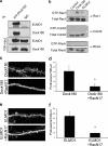
ELMO and Dock180 form a complex in hippocampal neurons and function through activating Rac1. a, co-immunoprecipitation of ELMO1 and Dock180 from hippocampal lysates. Hippocampal neurons (DIV 10) were lysed, and endogenous Dock180 was immunoprecipitated with a Dock180 antibody. The immunoprecipitated complex was analyzed by SDS-PAGE and immunoblotting with the indicated antibodies. IP, immunoprecipitation. b, cell lysates from hippocampal neurons infected with the Dock180- or Elmo1-overexpressing lentivirus were incubated with GST-PBD or GST-RBD, and bound small GTPases were detected with antibodies against Rac1, Cdc42, and RhoA. c, dominant negative Rac (RacN17) reverses the Dock180 overexpression phenotype. Hippocampal neurons were transfected with Dock180 and either an empty vector or RacN17 and imaged at DIV 17. d, quantification of protrusion density of neurons in c; *, p < 0.001 by Student's t test. e, dominant negative Rac (RacN17) reverses the ELMO1 overexpression phenotype. Hippocampal neurons were transfected with ELMO1 and either an empty vector or RacN17 and imaged at DIV 17. f, quantification of protrusion density of neurons in e; *, p < 0.001 by Student's t test.
FIGURE 5.
FIGURE 5.
RhoG is required for spine formation in cultured hippocampal neurons. a, expression of RhoG and ELMO1 in hippocampal neurons. Cultured hippocampal neurons were harvested at the indicated days in vitro, and the lysates were analyzed by Western blotting. b, co-immunoprecipitation of RhoG and ELMO1 from hippocampal lysates. Hippocampal neurons (DIV 10) were lysed, and endogenous RhoG was immunoprecipitated with a RhoG antibody. The immunoprecipitated complex was analyzed by SDS-PAGE and immunoblotting with the indicated antibodies. IP, immunoprecipitation. c, knockdown efficiency of RhoG shRNA. Cortical neurons were infected at DIV 7 with lentivirus expressing RhoG shRNA. Seventy-two hours after infection, neurons were lysed and analyzed by Western blotting. d, effects of RhoG knockdown on spine formation. Hippocampal neurons were transfected with either pSuper (Control) or pSuper-RhoG shRNA at DIV 6 and imaged at DIV 17. Venus was co-expressed to visualize spine morphology. Knockdown of RhoG significantly reduced the number of spines. e, quantification of protrusion density of neurons in b; *, p < 0.001 by Student's t test.
FIGURE 6.
FIGURE 6.
Activation of RhoG causes increased spine density and enlargement of the spine head. a, effects of overexpression of EGFP wild type RhoG, a dominant negative EGFP-RhoG T17N, and a constitutively active EGFP-RhoG Q61L on spine formation. Hippocampal neurons were transfected with pSuper, EGFP-RhoG Q61L, or EGFP-RhoG T17N at DIV 6 and imaged at DIV 19. b, quantification of protrusion density in neurons expressing EGFP-RhoG Q61L or EGFP-RhoG T17N. The values are means ± S.D. *, p < 0.001 by Student's t test. For each construct, images of 15–20 neurons from three independent culture preparations were analyzed. c, quantification of spine head size in neurons overexpressing RhoG Q61L. d, spines in RhoG Q61L-overexpressing neurons are apposed to presynaptic terminals. Hippocampal neurons expressing RhoG Q61L were immunostained with a Synapsin 1 antibody at DIV 17. IF, immunofluorescence.
FIGURE 7.
FIGURE 7.
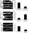
RhoG functions through the ELMO1/Dock180/Rac pathway. a, ELMO1 depletion reverses the RhoG Q61L overexpression phenotype. Hippocampal neurons were transfected with RhoG Q61L and either a control luciferase shRNA or ELMO1 shRNA at DIV 6 and imaged at DIV 17. b, quantification of protrusion density of neurons in a; *, p < 0.001 by Student's t test. c, ELMO1 overexpression rescues the RhoG T17N phenotype. Hippocampal neurons were transfected with RhoG T17N and either Venus (super-enhanced YFP) or ELMO1. Neurons were transfected at DIV 6 and imaged at DIV 17. d, quantification of protrusion density of neurons in c; *, p < 0.001 by Student's t test. e, dominant negative Rac (RacN17) reverses the RhoG Q61L overexpression phenotype. Hippocampal neurons were transfected with RhoG Q61L and either an empty vector or RacN17 and imaged at DIV 17. f, quantification of protrusion density of neurons in e; *, p < 0.001 by Student's t test.

References

    1. Hering H., Sheng M. (2001) Nat. Rev. Neurosci. 2, 880–888 - PubMed
    1. Sheng M., Hoogenraad C. C. (2007) Annu. Rev. Biochem. 76, 823–847 - PubMed
    1. Fiala J. C., Spacek J., Harris K. M. (2002) Brain Res. Brain Res. Rev. 39, 29–54 - PubMed
    1. Bonhoeffer T., Yuste R. (2002) Neuron 35, 1019–1027 - PubMed
    1. Cingolani L. A., Goda Y. (2008) Nat. Rev. Neurosci. 9, 344–356 - PubMed

Publication types

MeSH terms