

Quantifying the role of angiogenesis in malignant progression of gliomas: in silico modeling integrates imaging and histology
- PMID: 21900399
- PMCID: PMC3398690
- DOI: 10.1158/0008-5472.CAN-11-1399
Quantifying the role of angiogenesis in malignant progression of gliomas: in silico modeling integrates imaging and histology
Abstract
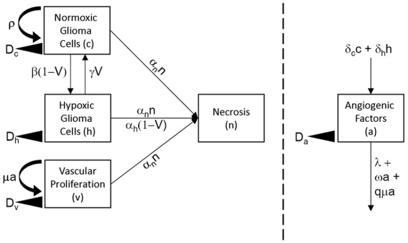
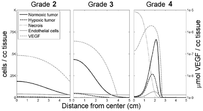
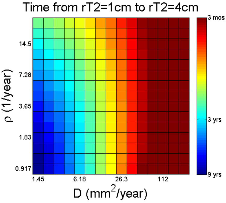
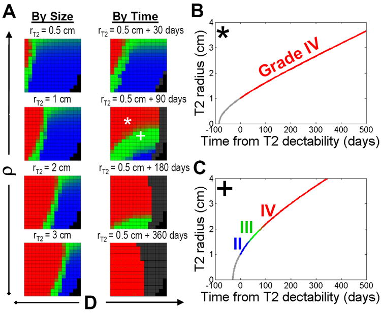
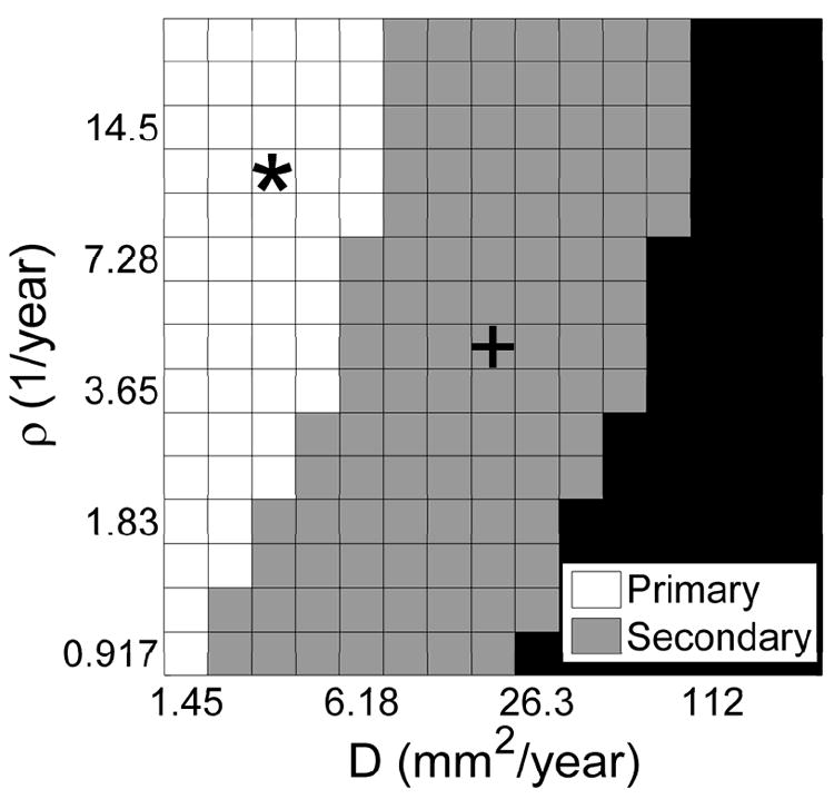
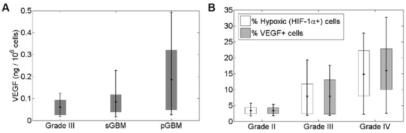
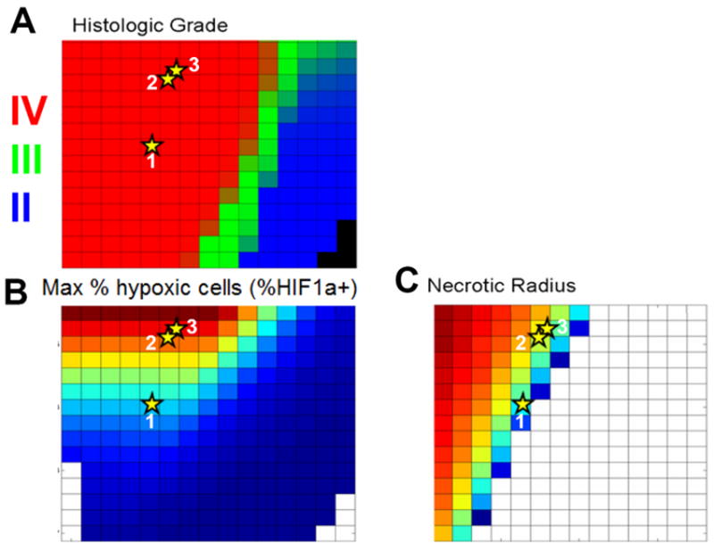
Gliomas are uniformly fatal forms of primary brain neoplasms that vary from low- to high-grade (glioblastoma). Whereas low-grade gliomas are weakly angiogenic, glioblastomas are among the most angiogenic tumors. Thus, interactions between glioma cells and their tissue microenvironment may play an important role in aggressive tumor formation and progression. To quantitatively explore how tumor cells interact with their tissue microenvironment, we incorporated the interactions of normoxic glioma cells, hypoxic glioma cells, vascular endothelial cells, diffusible angiogenic factors, and necrosis formation into a first-generation, biologically based mathematical model for glioma growth and invasion. Model simulations quantitatively described the spectrum of in vivo dynamics of gliomas visualized with medical imaging. Furthermore, we investigated how proliferation and dispersal of glioma cells combine to induce increasing degrees of cellularity, mitoses, hypoxia-induced neoangiogenesis and necrosis, features that characterize increasing degrees of "malignancy," and we found that changes in the net rates of proliferation (ρ) and invasion (D) are not always necessary for malignant progression. Thus, although other factors, including the accumulation of genetic mutations, can change cellular phenotype (e.g., proliferation and invasion rates), this study suggests that these are not required for malignant progression. Simulated results are placed in the context of the current clinical World Health Organization grading scheme for studying specific patient examples. This study suggests that through the application of the proposed model for tumor-microenvironment interactions, predictable patterns of dynamic changes in glioma histology distinct from changes in cellular phenotype (e.g., proliferation and invasion rates) may be identified, thus providing a powerful clinical tool.
Figures
References
-
- Brem S. The role of vascular proliferation in the growth of brain tumors. Clin Neurosurg. 1976;23:440–53. - PubMed
-
- Plate KH, Breier G, Weich HA, Mennel HD, Risau W. Vascular Endothelial Growth-Factor and Glioma Angiogenesis - Coordinate Induction of Vegf Receptors, Distribution of Vegf Protein and Possible in-Vivo Regulatory Mechanisms. Int J Cancer. 1994;59:520–9. - PubMed
-
- Harpold HL, Alvord EC, Jr, Swanson KR. The evolution of mathematical modeling of glioma proliferation and invasion. J Neuropathol Exp Neurol. 2007;66:1–9. - PubMed
-
- Swanson KR, Alvord EC, Jr, Murray JD. Virtual resection of gliomas: effects of location and extent of resection on recurrence. Mathematical and Computer Modeling. 2003;37:1177–90.
Publication types
MeSH terms
Grants and funding
LinkOut - more resources
Full Text Sources
Other Literature Sources
Medical
