. 2012 Jan 1;302(1):F103-15.
doi: 10.1152/ajprenal.00419.2011. Epub 2011 Sep 7.

WT1-interacting protein (Wtip) regulates podocyte phenotype by cell-cell and cell-matrix contact reorganization

Affiliations

WT1-interacting protein (Wtip) regulates podocyte phenotype by cell-cell and cell-matrix contact reorganization

Jane H Kim et al. Am J Physiol Renal Physiol. .

Abstract

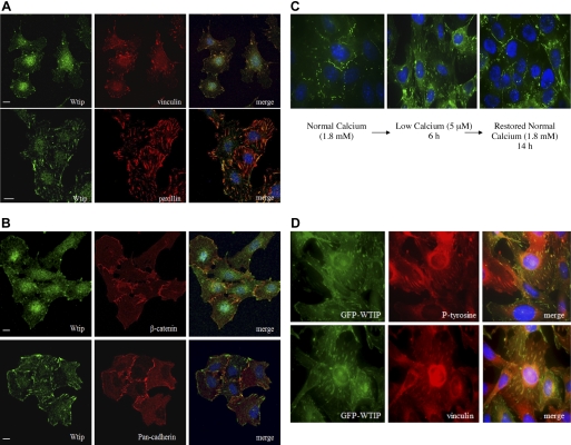
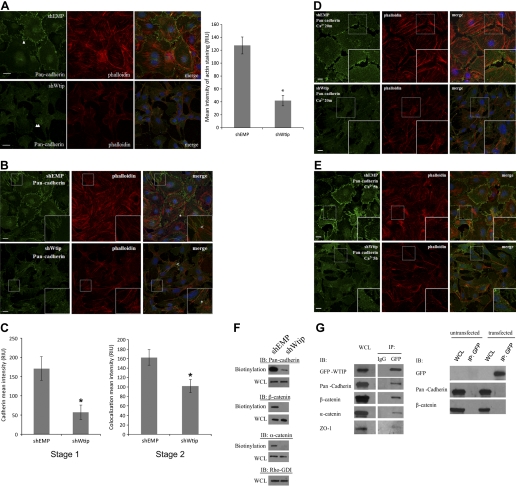
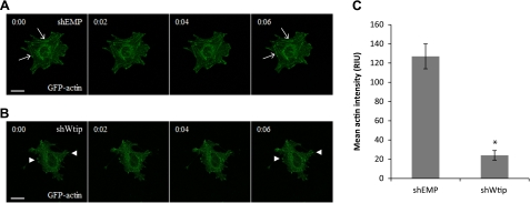
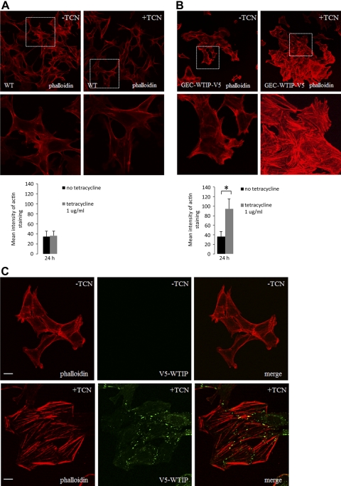
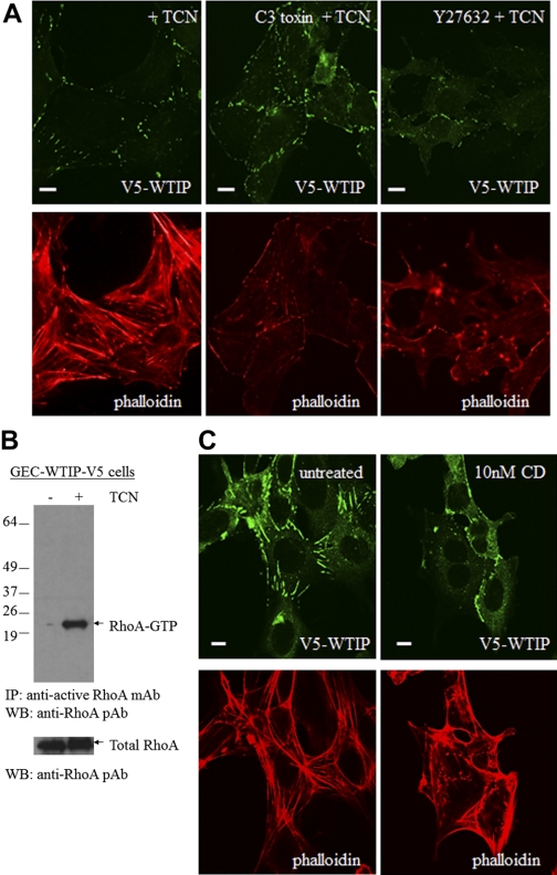
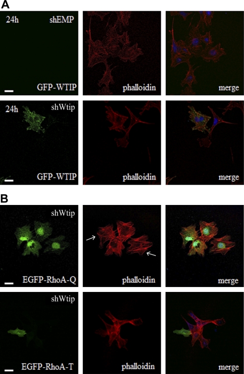
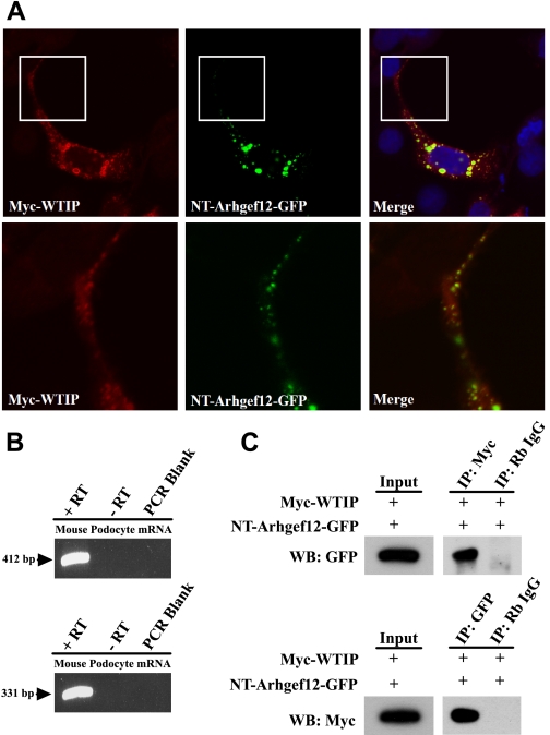
Podocytes respond to environmental cues by remodeling their slit diaphragms and cell-matrix adhesive junctions. Wt1-interacting protein (Wtip), an Ajuba family LIM domain scaffold protein expressed in the podocyte, coordinates cell adhesion changes and transcriptional responses to regulate podocyte phenotypic plasticity. We evaluated effects of Wtip on podocyte cell-cell and cell-matrix contact organization using gain-of- and loss-of-function methods. Endogenous Wtip targeted to focal adhesions in adherent but isolated podocytes and then shifted to adherens junctions after cells made stable, homotypic contacts. Podocytes with Wtip knockdown (shWtip) adhered but failed to spread normally. Noncontacted shWtip podocytes did not assemble actin stress fibers, and their focal adhesions failed to mature. As shWtip podocytes established cell-cell contacts, stable adherens junctions failed to form and F-actin structures were disordered. In shWtip cells, cadherin and β-catenin clustered in irregularly distributed spots that failed to laterally expand. Cell surface biotinylation showed diminished plasma membrane cadherin, β-catenin, and α-catenin in shWtip podocytes, although protein expression was similar in shWtip and control cells. Since normal actin dynamics are required for organization of adherens junctions and focal adhesions, we determined whether Wtip regulates F-actin assembly. Undifferentiated podocytes did not elaborate F-actin stress fibers, but when induced to overexpress WTIP, formed abundant stress fibers, a process blocked by the RhoA inhibitor C3 toxin and a RhoA kinase inhibitor. WTIP directly interacted with Rho guanine nucleotide exchange factor (GEF) 12 (Arhgef12), a RhoA-specific GEF enriched in the glomerulus. In conclusion, stable assembly of podocyte adherens junctions and cell-matrix contacts requires Wtip, a process that may be mediated by spatiotemporal regulation of RhoA activity through appropriate targeting of Arhgef12.


Figures

Fig. 1.
Fig. 1.
Endogenous WT1-interacting protein (Wtip) was localized to focal adhesions and cell-cell contacts in podocytes and is dynamically regulated by cell-cell adhesion. A: immunostaining with anti-Wtip (FITC), anti-vinculin, and anti-paxillin (TRITC). B: immunostaining for adherens junction proteins with anti-β-catenin and anti-pan-cadherin (TRITC). Nuclei were labeled with CY5-TOPRO-3. C: calcium switch assay demonstrated green fluorescent protein (GFP)-WTIP localization at cell-cell junctions in medium with normal calcium (NC) concentration (1.8 mM). With disruption of adhesion junctions in low-calcium (LC) medium (5 μM), GFP-WTIP localized in patches resembling focal adhesions (middle). GFP-WTIP retargeted to adherens junctions (left). D: in LC medium, GFP-WTIP colocalized with the focal adhesion markers phosphotyrosine and vinculin. Scale bars = 20 μm.
Fig. 2.
Fig. 2.
Wtip knockdown impaired stress fiber formation and dysregulated focal adhesion maturation. A: actin stress fibers were visualized by rhodamine-phalloidin staining in control vector (shEMP; top) and in shWtip (bottom) and costained with anti-WTIP (FITC). B: focal adhesions were immunostained with anti-vinculin (TRITC) in shEMP and shWtip. C: focal adhesion maturation was assessed by anti-vinculin immunostaining in shEMP and shWtip cells. Quantification of focal adhesion size was determined as described in experimental procedures. The bar graph shows mean relative intensity units for vinculin (right). D: shEMP and shWtip cells were immunostained with anti-paxillin, a marker of focal contacts. The bar graph shows mean relative intensity units for paxillin (right). E: phosphotyrosine levels of focal adhesions in shEMP and shWtip cells were assessed by immunostaining with PY99 as an indicator of focal adhesion maturation. The bar graph shows mean relative intensity units for phosphotyrosine (right). F: RT-PCR of shEMP and shWtip cells for Wtip, Limd1, and Gapdh transcripts. G: immunoblot analysis of shEMP (control) and shWtip (knockdown) podocytes for Wtip, paxillin, vinculin, focal adhesion kinase (FAK), and tubulin. Leica Quantify software was used for quantification. Scale bars = 20 μm. *P < 0.05.
Fig. 3.
Fig. 3.
Knockdown of Wtip affects proper cadherin junction assembly and targeting. shWtip and shEMP cells were stained with rhodamine-phalloidin and anti-pan-cadherin (A and B). Nuclei were labeled with CY5-TOPRO-3. Images in A show robust cadherin-based cell-cell contacts in shEMP podocytes (arrowhead) but not in shWtip cells (double arrowheads). The histograms quantify actin stress fiber abundance as described in experimental procedures. C: quantification of cell-cell adhesion formation in shEMP and Wtip knockdown cells as described in experimental procedures and results. shWtip podocytes are shown 20 min (D) and 5 h (E) after the switch from low to normal calcium. F: biotinylation demonstrated decreased plasma membrane localization of adherens junction proteins cadherin, β-catenin, and α-catenin to the membrane in shWtip vs. shEMP podocytes. Immunoblot (IB) analysis of whole cell lysates showed equal expression of cadherin, β-catenin, α-catenin, and RhoGDI. WCL, whole cell lysates. G: GFP-WTIP formed a complex with the indicated adherens junction proteins (left), but GFP failed to precipitate these proteins (right). Scale bars = 10 μm. *P < 0.05.
Fig. 4.
Fig. 4.
Wtip knockdown affects dynamic actin assembly. Time-lapse images of shEMP (A) and shWtip cells (B) transiently transfected with eGFP-actin (green) are shown. Arrows indicate stress fiber formation in shEMP cells, whereas arrowheads indicate actin clusters and the lack of actin stress fiber formation in shWtip cells. C: quantification of stress fiber formation in shEMP vs. shWtip cells (*P < 0.05). Scale bars = 20 μm.
Fig. 5.
Fig. 5.
Overexpression of GFP-WTIP enhances formation of actin stress fibers. A: F-actin assembly compared in control podocytes with and without tetracycline (TCN). Bar graph demonstrates mean actin staining using a single line scan across the nuclei of each (100 cells; n = 3 experiments). B: F-actin assembly in GEC-WTIP-V5 +/− TCN and quantified in the bar graph (*P ≤ 0.01, GEC-WTIP-V5 with TCN vs. no TCN). C: GEC-WTIP-V5 +/− TCN-assessed F-actin using rhodamine-phalloidin (left) and WTIP-V5 expression (FITC, middle). Merged images are shown on the right. Scale bars = 20 μm.
Fig. 6.
Fig. 6.
WTIP stress fiber formation was regulated through RhoA-dependent pathways. A: GEC-WTIP-V5 were preincubated with either a cell-permeable RhoA inhibitor, C3 toxin (top middle) or a Rho kinase inhibitor, Y27632 (top right), or were untreated (control) for 3 h followed by incubation with TCN for 24 h. B: RhoA activation assay in GEC-WTIP-V5 cells +/− TCN. C: low-dose cytochalasin D (CD; 10 nM) pretreatment of GEC-WTIP-V5 for 20 min followed by addition of TCN. Scale bars = 10 μm. IP, immunoprecipitated; WB, Western blotting.
Fig. 7.
Fig. 7.
Overexpression of GFP-WTIP or constitutively active RhoA rescued actin phenotype. A: transient transfection of an eGFP-WTIP expression vector demonstrated comparable actin stress fiber formation using rhodamine-phalloidin in shWtip and shEMP podocytes. B: transient transfection of EGFP-RhoAQ63L (RhoA-Q, constitutively active) and EGFP- RhoAT19N (RhoA-T, dominant negative) constructs into shWtip cells demonstrated RhoA activity is required for stress fiber formation. Arrows indicate actin stress fiber tips. Nuclei were labeled with CY5-TOPRO-3.
Fig. 8.
Fig. 8.
Myc-WTIP interacted with the PDZ domain-containing, amino terminal (NT) of the RhoA GEF, Arhgef12. A: COS7 cells were cotransfected with Myc WTIP and NT-Arhgef12-GFP constructs. Immunofluorescence labeling was done with rabbit polyclonal anti-Myc antibody followed by Alexa Fluor 568-conjugated anti-rabbit secondary antibody. Myc-WTIP (red) expression colocalizes with Arhgef12 (green) in the merged image. Magnified view of selected areas from top show colocalization in cell extension. B: RT-PCR was done in mouse podocytes using two primer sets amplifying different regions of Arhgef12 message. PCR products of expected size (412 and 331 bp) were observed for the N-terminal PDZ domain-containing and internal coding region of Arhgef12, respectively. C: coimmunoprecipitation was done in 293 cells transfected with Myc-WTIP and Arhgef12-GFP. Cell lysates were immunoprecipitated using anti-Myc or anti-GFP antibodies, and the immunoprecipitates were examined by Western blotting using anti-GFP or anti-Myc antibodies, respectively. Rabbit IgG was used as a control for immunoprecipitation. Input represented 5% of cell lysates used in the coimmunoprecipitation.

References

    1. Adams CL, Chen YT, Smith SJ, James Nelson W. Mechanisms of epithelial cell-cell adhesion and cell compaction revealed by high-resolution tracking of E-cadherin-green fluorescent protein. J Cell Biol 142: 1105–1119, 1998 - PMC - PubMed
    1. Amano M, Chihara K, Kimura K, Fukata Y, Nakamura N, Matsuura Y, Kaibuchi K. Formation of actin stress fibers and focal adhesions enhanced by Rho-kinase. Science 275: 1308–1311, 1997 - PubMed
    1. Bhadriraju K, Yang M, Alom Ruiz S, Pirone D, Tan J, Chen CS. Activation of ROCK by RhoA is regulated by cell adhesion, shape, and cytoskeletal tension. Exp Cell Res 313: 3616–3623, 2007 - PMC - PubMed
    1. Craig SW, Johnson RP. Assembly of focal adhesions: progress, paradigms, and portents. Curr Opin Cell Biol 8: 74–85, 1996 - PubMed
    1. Dawid IB, Breen JJ, Toyama R. LIM domains: multiple roles as adapters and functional modifiers in protein interactions. Trends Genet 14: 156–162, 1998 - PubMed

Publication types

MeSH terms