. 2011 Nov;22(21):4079-92.
doi: 10.1091/mbc.E11-02-0121. Epub 2011 Sep 7.

Membrane-targeted WAVE mediates photoreceptor axon targeting in the absence of the WAVE complex in Drosophila

Affiliations

Membrane-targeted WAVE mediates photoreceptor axon targeting in the absence of the WAVE complex in Drosophila

Raiko Stephan et al. Mol Biol Cell. 2011 Nov.

Abstract

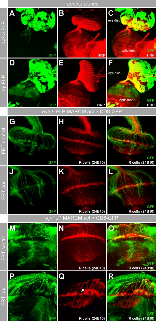
A tight spatial-temporal coordination of F-actin dynamics is crucial for a large variety of cellular processes that shape cells. The Abelson interactor (Abi) has a conserved role in Arp2/3-dependent actin polymerization, regulating Wiskott-Aldrich syndrome protein (WASP) and WASP family verprolin-homologous protein (WAVE). In this paper, we report that Abi exerts nonautonomous control of photoreceptor axon targeting in the Drosophila visual system through WAVE. In abi mutants, WAVE is unstable but restored by reexpression of Abi, confirming that Abi controls the integrity of the WAVE complex in vivo. Remarkably, expression of a membrane-tethered WAVE protein rescues the axonal projection defects of abi mutants in the absence of the other subunits of the WAVE complex, whereas cytoplasmic WAVE only slightly affects the abi mutant phenotype. Thus complex formation not only stabilizes WAVE, but also provides further membrane-recruiting signals, resulting in an activation of WAVE.


Figures

FIGURE 1:
FIGURE 1:
abi is required for photoreceptor targeting. (A) Schematic overview of the abi locus with the neighboring twf gene. The position of the transposon used to generate the abi mutant is indicated. A dashed line indicates the extension of the genomic deletion in the abi mutant. (B and C) Western blot analysis of larval brain extracts. (B) The abiΔ20 mutation results in a loss of Abi protein, whereas (C) the expression of the adjacent Twf protein is unaffected. (D–P) Analysis of photoreceptor projection pattern in the fly larval visual system in abi mutants. (D) Schematic overview of the projection pattern of photoreceptor axons (adapted from Tayler and Garrity, 2003). (E) In wild type, Abi (gray) is expressed in photoreceptor axons, as well as in the lamina (la, arrows) and medulla (me, asterisk). (F–H) R-cell axons (green, R1–R8; antibody 24B10) show a stereotyped projection to the lamina and medulla with R2–R5 (red, marked by rough-τlacZ; α-β-galactosidase) terminating in the lamina (note a few axons overshooting the lamina, asterisk in G). (J–M) The loss of abi leads to a highly abnormal targeting of R-cell axons with axonal bundling and uneven appearance of the lamina with gaps and clumps (K). The vast majority of the R2–R5 axons fail to terminate in the lamina and terminate in deeper layer medulla (L). (I and N) Rescue of the abi-dependent projection defects of R-cell axons. Neural resupply (N) of Abi (elavGal4 > Abi) rescues abi-mediated targeting abnormalities of R-cell axon whereas (I) glial reexpression of Abi does not (repoGal4 > Abi). (O–P) Quantification of the photoreceptor targeting defects. (O) The number of axonal bundles in the medulla per optic lobe was quantified for the indicated genotypes. abi, elav > Abi: neural reexpression of Abi in abi mutant animals. abi, repo > Abi: glial reexpression of Abi in abi mutant animals. Error bars represent SEM. ***, p < 0.001 (analysis of variance [ANOVA]); n.s., not significant. (P) Severity of R2–R5 overshooting defects in the indicated genotypes. Animals used for the quantification of axonal bundles were grouped according to the numbers of R2–R5 axons overshooting the lamina. The numbers represent percent of optic lobes with R2–R5 axons in the medulla. abi, elav > Abi: neural reexpression of Abi in abi mutant animals.
FIGURE 2:
FIGURE 2:
abi function is required in target area neurons. (A–F) Comparison of the spatial activity of different flippase sources used for clonal analysis. (A–C) Eye disk–specific flippase activity (ey3.5-FLP) induces dominantly CD8-GFP–labeled cell clones (green) only in the eye imaginal disk including R-cells and their axons. (D–F) Genetic mosaics induced by ey-FLP leads to CD8-GFP expression in the eye disk as well as in the brain target area. Anti-HRP (red) was used as general neuronal marker to visualize brain structures. (G–R) MARCM analysis of abi in the developing visual system. R-cell axons are visualized with the antibody 24B10 (red); cell clones are dominantly marked by CD8-GFP expression (green). (G–L) Analysis of abi mutant cell clones generated only in the eye disk by ey3.5-FLP. As in control clones (G–I), axons of abi mutant R-cells show a normal targeting (J–L). (M–R) Analysis of abi mutant cell clones generated in the eye disk and the brain target area by ey-FLP. In contrast to control clones (M–O), mutant cell clones lacking abi function (P–R) are associated with severe lamina targeting defects (arrow in Q).
FIGURE 3:
FIGURE 3:
abi controls the organization of an axonal scaffold in the target area. (A–H) Analysis of scaffold axon fascicles in the brain target area. (A, C, E, and G) In wild-type scaffold, axon fascicles grow out from two Wingless (Wg)-expressing domains (green, marked by wg-lacZ; visualized with α-β-galactosidase). The axon fascicles extend to neuropile regions marked by intense horseradish peroxidase staining (red). Glia cell nuclei (blue, anti-Repo) are located around the neuropile regions. (B, D, F, and H) In abi mutant brains the organization of the brain target area in the visual system is disturbed. The Wg-expressing domains appear broadened and irregular (arrows in B). The trajectories of wg-lacZ–labeled axon fascicles are abnormal. The neuropile regions and the position of the surrounding glia cell nuclei also appear disturbed (compare D vs. C and F vs. E).
FIGURE 4:
FIGURE 4:
The N-terminus of Abi is required during photoreceptor targeting. (A) Schematic overview of the domain structure of Abi and Abi variants used in the analysis. Numbers indicate the amino acid position of an indicated domain. WAB (red); HHR (gray) required for Kette binding; P (gray): proline-rich regions; SH3 (magenta) required for WASP interaction. rescue: summary of ability of Abi variants to rescue R-cell targeting defect upon neural reexpression (+ indicates rescue; − indicates no rescue). (B–E′′) Representative images of projection patterns of all R-cell axons and R2–R5 axons of the indicated genotypes. R-cell axons (R1–R8, red) are visualized by anti-24B10, and R2–R5 axons (green) are marked by the ro-τlacZ reporter (α-β-galactosidase). Abi variants were expressed in all neurons using the elavGal4 driver line. Whereas full-length Abi and a C-terminal truncated Abi protein (AbiΔC) (B–C′′) are able to rescue the abi-dependent projection defects, an Abi protein lacking the N-terminal WAB domain (AbiΔN) fails to rescue the R-cell targeting phenotype (D–D′′). Expression of the Abi N-terminus alone (Abi-WAB-HHR) also restores the projection defects of R-cell axons. *RFP expression in the Bolwig's nerve marks the 68E landing site of the ΦC31 integrase system for generating site-specific transgenic fly lines (Bischof et al., 2007). (F–G) Quantification of the rescue experiments using Abi variants. (F) The number of axonal bundles in the medulla per optic lobe was quantified for the indicated Abi variants upon neural reexpression in the abi mutant background. Abi: full-length Abi; AbiΔC: Abi variant lacking C-terminal SH3 domain; AbiΔN: Abi variant lacking N-terminal WAB domain; Abi-WAB-HHR: Abi variant consisting only of N-terminal WAB- and HHR domain. Error bars represent SEM and data sets of rescue experiments refer to the abi mutant. ***, p < 0.001 (ANOVA); n.s., not significant. (G) Severity of R2–R5 overshooting defects in the indicated genotypes. Numbers represent percent of optic lobes with R2–R5 axons in the medulla.
FIGURE 5:
FIGURE 5:
Abi acts through WAVE during photoreceptor axon targeting. (A–G) Representative images of R-cell projection patterns of the indicated genotypes. R-cell axons (green) are visualized with 24B10. (A–C) Compared with wild type, the loss of wave, but not wasp, leads to an axonal targeting phenotype similar to abi mutants with axonal bundling and uneven appearance of the lamina with gaps and clumps. (D–G) Rescue of the wave-dependent projection defect. Resupply of full-length Wave in glial cells (repoGal4 > WAVE) does not improve the R-cell targeting in the wave mutant background (D). Neural reexpression of full-length Wave (elavGal4 > WAVE) in wave mutants restores a wild-type appearance of the projection pattern of R-cell axons (E). In contrast, a Wave variant lacking the N-terminal Abi-binding region (WAVEΔN) is unable to rescue the targeting defects neurally expressed in wave mutants (F). Expression of a Wave protein missing the basic region (WAVEΔB) mediating membrane association only partially restores the wave-dependent targeting defects upon neural expression (G). (H) Schematic overview of the domain structure of WAVE and WAVE variants used in the rescue experiments. WHD (green): WAVE homology domain mediating Abi binding; B (gray): basic region required for lipid binding; PolyPro (blue): proline-rich regions; VCA (red) region required for Arp2/3 binding and activation. rescue: summary of ability of WAVE variants to rescue R-cell targeting defect upon neural reexpression (+ indicates full rescue; − indicates no rescue; −/+ indicates partial rescue).
FIGURE 6:
FIGURE 6:
Members of the WAVE complex are required for the integrity of WAVE in vivo. (A) Gel filtration profiles of WASP, WAVE, Abi, and Kette from lysates of heads of adult flies. The elution profile of proteins of known molecular weight is indicated at the bottom. Abi mainly cofractionates with WAVE and Kette (fractions 18–20). A portion of Abi cofractionates also with WASP (fractions 21–22) that hardly contain WAVE and Kette. (B–C) WAVE stability requires abi function in vivo. Cell clones lacking abi were generated in the eye imaginal disk and are marked by the absence of GFP (green, GMR-GFPMyr). Mutant cell clones (dashed lines in C) in the eye imaginal disk lacking abi function show a pronounced instability of endogenous WAVE protein (red, anti-WAVE). (D–E) Western blot analysis of larval brain extracts from wild type, kette, abi, and mutant larvae expressing indicated proteins. Quantification of protein levels (E) was done with the Image QuantTL software package (GE Healthcare, Waukesha, WI). Data were normalized with respect to tubulin levels. The loss of abi or kette leads to an instability of the members of the WAVE complex (WAVE, Abi, Kette, Sra-1) in vivo. Concomitantly, protein levels of WASP appear unchanged. Neural expression of Abi and Kette in abi and kette mutants, respectively, restores the integrity of the WAVE complex in vivo. Expression of an N-terminal truncated Abi protein does not lead to an increase of WAVE levels in the mutant background (blue in (E), quantification for lane 6 vs. lane 4). In contrast, an Abi variant lacking the WASP-binding domain (AbiΔC) elevates WAVE protein levels upon neural expression in the mutant background (blue in (E), quantification for lane 7 vs. lane 4). Please note that the used anti-Abi antibody does not recognize the AbiΔC protein variant (see Supplemental Information).
FIGURE 7:
FIGURE 7:
WAVE activity at the membrane in the absence of the WAVE complex (A–F) Analysis of ability of WAVE variants to rescue the abi-dependent projection defects. (A–D′′) Representative images of projection patterns of all R-cell axons and R2–R5 axons of the indicated genotypes. R-cell axons (R1–R8, red) are visualized by anti-24B10 and R2–R5 axons (green) are marked by the ro-τlacZ reporter (α-β-Galactosidase). WAVE variants were expressed in all neurons using the elavGal4 driver line. A full-length WAVE protein expressed in neurons in the abi mutant background weakly rescues the abi-dependent projection defects (A–A′′). A membrane-targeted WAVE variant expressed from an arbitrary genomic site (Myr-WAVE, B–B′′), as well as expressed from a specific site (Myr-WAVE68E, C–C′′) partially rescues the projection defects by abi. In contrast, a membrane-targeted Wave lacking the Arp2/3-activating VCA domain (Myr-WAVEΔVCA68E, D–D′′) expressed from the same genomic site fails completely to rescue the abi mutant phenotype. * RFP expression in the Bolwig's nerve marks the 68E landing site of the ΦC31 integrase system for generating site-specific transgenic fly lines (Bischof et al., 2007). (E–F) Quantification of the rescue experiments using WAVE variants in the abi mutant background. (E) The number of axonal bundles in the medulla per optic lobe was quantified for the indicated WAVE variants upon neural reexpression in the abi mutant background. WAVE: full-length WAVE; Myr-WAVE: membrane-tethered WAVE expressed from an arbitrary genomic site; Myr-WAVE68E: membrane-tethered WAVE expressed from a specific site (68E); Myr-WAVEΔVCA68E: membrane-tethered WAVE lacking the Arp2/3-activating domain expressed from a specific site (68E). Error bars represent SEM and data sets of rescue experiments were referred to the abi mutant. ***, p < 0.001; **, p < 0.01 (ANOVA); n.s., not significant. (F) Severity of R2–R5 overshooting defects in the indicated genotypes. Numbers represent percentage of optic lobes with R2–R5 axons in the medulla.

Similar articles

Cited by

References

    1. Anton IM, Jones GE, Wandosell F, Geha R, Ramesh N. WASP-interacting protein (WIP): working in polymerisation and much more. Trends Cell Biol. 2007;17:555–562. - PubMed
    1. Bazigou E, Apitz H, Johansson J, Loren CE, Hirst EMA, Chen PL, Palmer RH, Salecker I. Anterograde jelly belly and Alk receptor tyrosine kinase signaling mediates retinal axon targeting in Drosophila. Cell. 2007;128:961–975. - PubMed
    1. Ben-Yaacov S, Le Borgne R, Abramson I, Schweisguth F, Schejter ED. Wasp, the Drosophila Wiskott-Aldrich syndrome gene homologue, is required for cell fate decisions mediated by Notch signaling. J Cell Biol. 2001;152:1–13. - PMC - PubMed
    1. Berger J, Senti KA, Senti G, Newsome TP, Asling B, Dickson BJ, Suzuki T. Systematic identification of genes that regulate neuronal wiring in the Drosophila visual system. PLoS Genet. 2008;4:e1000085. - PMC - PubMed
    1. Bischof J, Maeda RK, Hediger M, Karch F, Basler K. An optimized transgenesis system for Drosophila using germ-line-specific phiC31 integrases. Proc Natl Acad Sci USA. 2007;104:3312–3317. - PMC - PubMed

Publication types

MeSH terms

Substances

LinkOut - more resources