. 2011 Dec;106(6):2876-87.
doi: 10.1152/jn.00312.2011. Epub 2011 Sep 7.

Astrocytes promote peripheral nerve injury-induced reactive synaptogenesis in the neonatal CNS

Affiliations

Astrocytes promote peripheral nerve injury-induced reactive synaptogenesis in the neonatal CNS

Fu-Sun Lo et al. J Neurophysiol. 2011 Dec.

Abstract

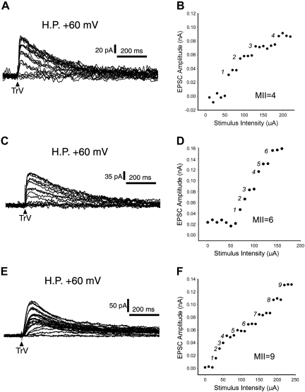
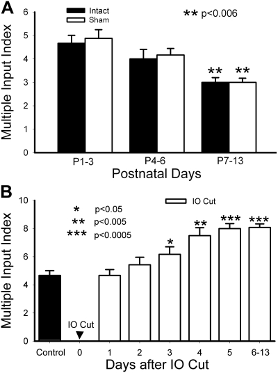
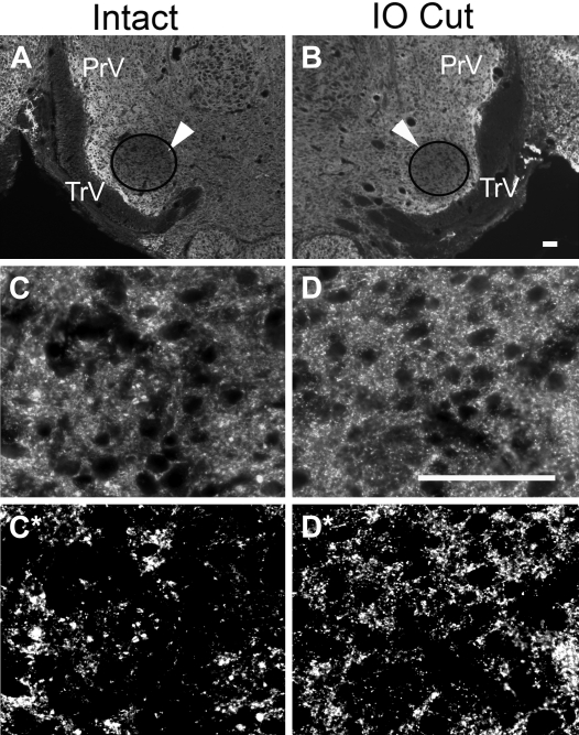
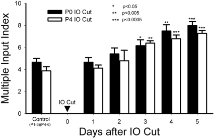
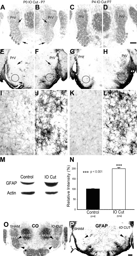
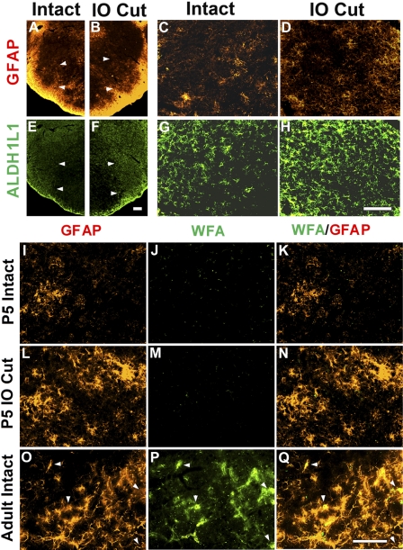
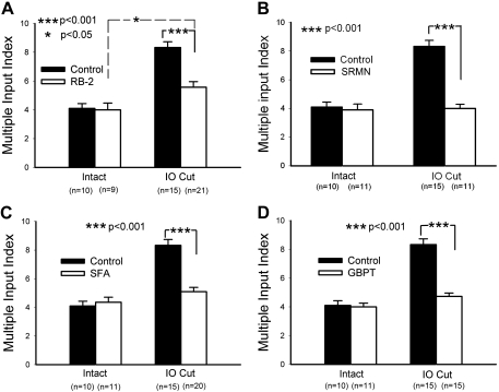
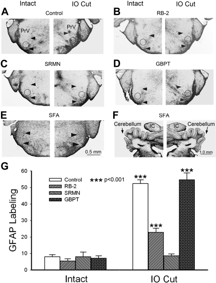
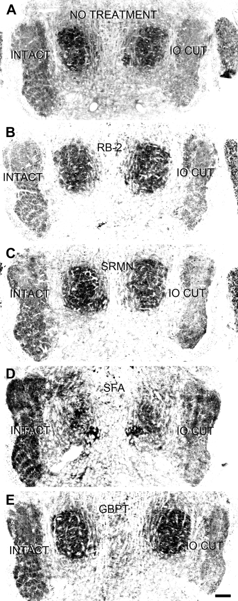
Neonatal damage to the trigeminal nerve leads to "reactive synaptogenesis" in the brain stem sensory trigeminal nuclei. In vitro models of brain injury-induced synaptogenesis have implicated an important role for astrocytes. In this study we tested the role of astrocyte function in reactive synaptogenesis in the trigeminal principal nucleus (PrV) of neonatal rats following unilateral transection of the infraorbital (IO) branch of the trigeminal nerve. We used electrophysiological multiple input index analysis (MII) to estimate the number of central trigeminal afferent fibers that converge onto single barrelette neurons. In the developing PrV, about 30% of afferent connections are eliminated within 2 postnatal weeks. After neonatal IO nerve damage, multiple trigeminal inputs (2.7 times that of the normal inputs) converge on single barrelette cells within 3-5 days; they remain stable up to the second postnatal week. Astrocyte proliferation and upregulation of astrocyte-specific proteins (GFAP and ALDH1L1) accompany reactive synaptogenesis in the IO nerve projection zone of the PrV. Pharmacological blockade of astrocyte function, purinergic receptors, and thrombospondins significantly reduced or eliminated reactive synaptogenesis without changing the MII in the intact PrV. GFAP immunohistochemistry further supported these electrophysiological results. We conclude that immature astrocytes, purinergic receptors, and thrombospondins play an important role in reactive synaptogenesis in the peripherally deafferented neonatal PrV.


Figures

Fig. 1.
Fig. 1.
Examples of multiple input index (MII). A, C, and E: example records of multiple innervation of barrelette neurons. Superimposed excitatory postsynaptic currents (EPSCs) are shown at a holding potential (HP) of +60 mV along with progressively increasing stimulus intensity from 0 to 350 μA at steps of 10 μA. Note that the EPSC amplitudes fluctuate in a stepwise manner. B, D, and F: plots of EPSC amplitude against stimulus intensity. The amplitudes show abrupt jumps as the stimulus intensity increases. TrV, trigeminal tract.
Fig. 2.
Fig. 2.
MII of barrelette neurons in the trigeminal principal nucleus (PrV). A: the MII of single barrelette neurons declined during postnatal development in both intact (filled bars) and sham-operated PrV (open bars). Note that there is no difference between intact and sham-operated PrV. The MII on postnatal days 7–13 (P7–P13) is significantly (P < 0.006) smaller than that on P1–P3. B: time course of synaptogenesis after deafferentation. One day after deafferentation [infraorbital (IO) cut], the MII is the same as that in intact PrV on P1–P3 (control). The MII begins to increase from 2 days after IO cut and reaches a plateau after 5 days later. Compared with 1 day postdeafferentation, the increase in the MII is significant on 3–5 days postdeafferentation (*P < 0.05; **P < 0.005; ***P < 0.0005).
Fig. 3.
Fig. 3.
Synaptophysin immunohistochemistry. A and B: low-power view of synaptophysin immunohistochemistry in the intact (A) and deafferented PrV (B). Arrowheads point to the regions of interest (ROI) over the barrelette region of the PrV from where the micrographs were taken at higher magnification. C and D: higher magnification views from intact (C) and deafferented sides (D) show the density of synaptic puncta in deafferented PrV under fluorescence. C* and D* show the same fields after thresholding (see materials and methods) for intensity measurements with the NIS Elements program. The density of the fluorescence is higher than that in the intact PrV. Scale bar, 100 μm.
Fig. 4.
Fig. 4.
Time course of reactive synaptogenesis after IO cut on P0 and P4 (during and after the critical period for structural plasticity). The MII of the first day after IO cut on P0 (filled bar) and on P4 (open bar) is similar to the control value. Compared with 1 day postdeafferentation, the increase in the MII is significant on 3–5 days postdeafferentation in both cases (*P < 0.05; **P < 0.005; ***P < 0.0005). These results indicate that IO nerve injury-induced reactive synaptogenesis is not confined to the critical period.
Fig. 5.
Fig. 5.
Effects of P0 and P4 IO nerve cut on the PrV and astrocytosis. A: cytochrome oxidase (CO) histochemistry revealed the barrelette patterns in the intact control side at P7. Arrows point to the IO nerve-recipient zone of the PrV in this and other micrographs. Whisker-specific barrelette rows (a–e) are indicated. B: the deafferented side of the same section shown in A. Note that the barrelettes have dissolved or did not form. C and D: CO staining in the intact (C) and deafferented sides (D) of the same P7 brain stem from a pup that underwent IO cut on P4 (after the critical period). Note that 5 rows of barrelettes are present in the deafferented PrV. E: glial fibrillary acidic protein (GFAP) labeling in the control PrV at P5. F: marked astrocytosis and high levels of GFAP expression in the IO nerve-recipient zone of the PrV in the deafferented side 4 days after P0 IO cut. Intense astrocytosis and GFAP staining is also visible after the critical period. G and H: control (G) and deafferented sides (H) of the same P7 brain stem after P4 IO cut. Note the high level of GFAP immunostaining in the IO nerve-recipient zone of the deafferented PrV (H). Higher magnification views of GFAP immunostaining are shown in I–L; these micrographs are from ROIs (marked by circles) of the PrV of the same sections shown in E–H. Scale bar in D is 200 μm for A–H and 20 μm for I–L. M: Western blots of GFAP expression on the ventral PrV (barrelette region) of P5 rat pups. N: the relative intensity of GFAP in the deafferented PrV was significantly higher (***P < 0.001) than that in the intact PrV. O: sham operation did not change barrelette patterns. P: sham operation did not upregulate GFAP expression. Scale bar, 100 μm.
Fig. 6.
Fig. 6.
Expression of glial markers and chondroitin sulfate proteoglycans (CSPGs). Top: both GFAP and ALDH1L1 expression are upregulated in the deafferented PrV. Low- and high-power photomicrographs of the intact PrV (A and C) show low GFAP expression, whereas in the deafferented PrV (B and D) GFAP expression is upregulated. Similarly, ALDH1L1 expression is upregulated in the denervated PrV (F and H) compared with the intact side (E and G). Note that ALDH1L1 labeled more astrocytes than GFAP, indicating that it is a better pan-astrocyte marker. Bottom micrographs illustrate the absence of Wisteria floribunda agglutinin (WFA) staining for CSPGs in the neonatal PrV (J and M), whereas the same procedure shows high levels of CSPG staining in the adult PrV (P). The same sections were immunostained with GFAP (I, L, and O), and double labeling is shown in K, N, and Q. Arrowheads point to astrocytes double-labeled with both WFA and GFAP. Scale bar, 100 μm.
Fig. 7.
Fig. 7.
MII analysis after various drug treatments in the intact (control) and deafferented (IO cut) PrV. A: application of reactive blue-2 (RB-2) partially blocked reactive synaptogenesis (IO cut, filled vs. open bars) without affecting developmental synaptogenesis in the control side (intact, filled vs. open bars). B: application of suramin (SRMN), a broad-spectrum antagonist for P2 class receptors, completely blocked the reactive synaptogenesis (IO cut, filled vs. open bars) with no effects on the control side (intact, filled vs. open bars). C: application of sodium fluoroacetate (SFA), an inhibitor of astrocyte metabolism, completely blocked the reactive synaptogenesis in the IO cut side (filled vs. open bars) but did not affect the control side (filled vs. open bars). D: application of gabapentin (GBPT), an antagonist for thrombospondin (TSP) receptor, also completely blocked the reactive synaptogenesis (IO cut, filled vs. open bars), yet it did not affect the MII in the control side (intact, filled vs. open bars). *P < 0.05; ***P < 0.001.
Fig. 8.
Fig. 8.
Analysis of GFAP expression after various drug treatments in the intact (control) and deafferented (IO cut) PrV. A: example micrographs of a control animal showing higher GFAP labeling in the ROI (circles) of the barrelette region (arrowheads) in the deafferented (IO cut) PrV than in the intact PrV. B: after RB-2 injections, the GFAP labeling in the deafferented side was less than that in control animals, but the labeling in the intact side was not changed. C: after SRMN injections, the GFAP labeling in the deafferented side was similar to that of the intact side, which was also similar to that of control PrV. D: after GBPT injections, the GFAP labeling in both sides was similar to that of controls, suggesting GBPT does not alter GFAP expression. E: application of SFA led to a conspicuous increase in GFAP labeling all over the brain stem. F: upregulation of GFAP expression was also seen in the cerebellum and inferior colliculus (IC). G: averaged measurements of GFAP labeling show that drug treatments did not affect GFAP expression in the intact PrV. In the deafferented PrV, RB-2 decreased GFAP labeling, SRMN completely blocked the increased GFAP labeling, and GBPT had no effect on GFAP labeling. ANOVA showed that only control, RB-2, and GBPT values are higher (***P < 0.001) than other values.
Fig. 9.
Fig. 9.
Pharmacological interventions aimed at perturbing astrocyte function do not interfere with formation and maintenance of barrelette patterns in the intact PrV. A comparison of intact and IO cut sides in the same brains from P5 rat pups that underwent P0 IO damage and includes images of controls (no drug treatment; A) and pups injected with RB-2 (B), SRMN (C), SFA (D), and GBPT (E). Note that in all cases the intact side has barrelette patterns, whereas the IO cut side has no patterns. Scale bar, 100 μm.

Similar articles

Cited by

References

    1. Abbracchio MP, Burnstock G, Boeynaems JM, Barnard EA, Boyer JL, Kennedy C, Knight GE, Fumagalli M, Gachet C, Jacobson KA, Weisman GA. International Union of Pharmacology LVIII: update on the P2Y G protein-coupled nucleotide receptors: from molecular mechanisms and pathophysiology to therapy. Pharmacol Rev 58: 281–341, 2006 - PMC - PubMed
    1. Adams JC. Thrombospondins: multifunctional regulators of cell interactions. Annu Rev Cell Dev Biol 17: 25–51, 2001 - PubMed
    1. Allen NJ, Barres BA. Signaling between glia and neurons: focus on synaptic plasticity. Curr Opin Neurobiol 15: 542–548, 2005 - PubMed
    1. Andersson M, Blomstrand F, Hanse E. Astrocytes play a critical role in transient heterosynaptic depression in the rat hippocampal CA1 region. J Physiol 585: 843–852, 2007 - PMC - PubMed
    1. Arsenault D, Zhang ZW. Developmental remodeling of the lemniscal synapses in the ventral basal thalamus of the mouse. J Physiol 573: 121–132, 2006 - PMC - PubMed

Publication types

MeSH terms