. 2011 Nov;18(11):1823-33.
doi: 10.1128/CVI.05262-11. Epub 2011 Sep 7.

Immunization of mice with Lactobacillus casei expressing a beta-intimin fragment reduces intestinal colonization by Citrobacter rodentium

Affiliations

Immunization of mice with Lactobacillus casei expressing a beta-intimin fragment reduces intestinal colonization by Citrobacter rodentium

P C D Ferreira et al. Clin Vaccine Immunol. 2011 Nov.

Abstract

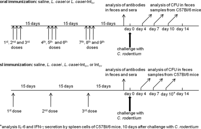
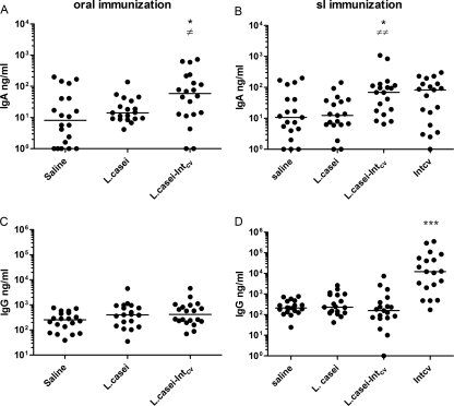
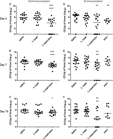
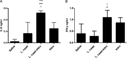
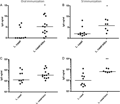
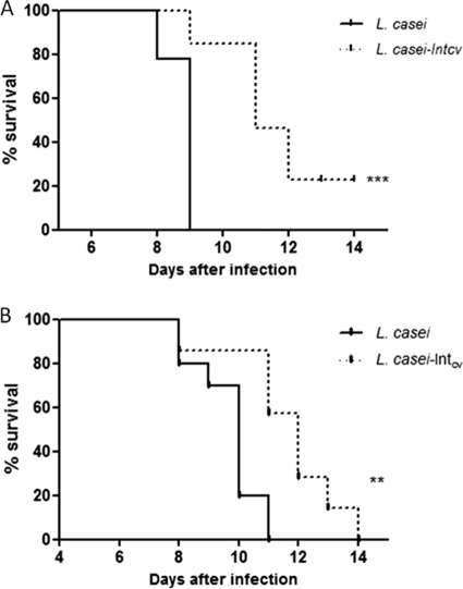
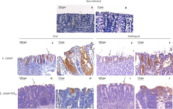
Enteropathogenic Escherichia coli (EPEC) is a common cause of diarrhea in children from developing countries. Intimate adhesion of the bacteria to intestinal cells occurs via binding of the adhesin intimin to the TIR receptor exposed on cell surfaces. Here, Lactobacillus casei expressing a fragment of β-intimin (L. casei-Int(cv)) was tested as mucosal vaccines in mice against intestinal colonization with the murine pathogen Citrobacter rodentium. Oral or sublingual immunization of C57BL/6 mice with L. casei-Int(cv) induced anti-Int(cv) IgA in feces but no IgG in sera. Conversely, anti-Int(cv) IgG was induced in the sera of mice after sublingual immunization with purified Int(cv). All vaccines were able to decrease C. rodentium recovery from feces. However, this reduction was more evident and sustained over time in mice immunized with L. casei-Int(cv) by the sublingual route. These mice also displayed an increase in interleukin 6 (IL-6) and gamma interferon (IFN-γ) secretion by spleen cells 10 days after infection. Additionally, oral or sublingual immunization of C3H/HePas mice, which are highly susceptible to C. rodentium infection, with L. casei-Int(cv) induced anti-Int(cv) antibodies and significantly increased survival after challenge. Immunohistological analysis of colon sections revealed that C. rodentium was located in deep fractions of the tissue from C3H/HePas mice immunized with L. casei whereas superficial staining was observed in colon sections from mice immunized with L. casei-Int(cv.) The results indicate that vaccines composed of L. casei expressing intimin may represent a promising approach and that the C3H/HePas infection model with C. rodentium can be used to evaluate potential vaccines against EPEC.


Figures

Fig. 1.
Fig. 1.
Immunization and challenge of C57BL/6 and C3H/HePas mice with C. rodentium. Oral immunization was performed using a gavage. Mice received saline, L. casei (1010 CFU), or L. casei-Intcv (1010 CFU) on three consecutive days at 15-day intervals, totaling 9 doses. S.l. immunization was performed in three doses of saline, L. casei (109 CFU), L. casei-Intcv (109 CFU), or Intcv (15 μg). Fifteen days after the last immunization, the mice were challenged with 5 × 10⁸ CFU of C. rodentium by the oral route, and colonization or survival was followed in C57BL/6 or C3H/HePas mice, respectively. Anti-Intcv antibodies were measured in blood and feces before challenge or in feces at different periods after challenge. Secretion of cytokines by spleen cells collected at the endpoint of the colonization experiment (10 days after challenge) was measured in C57BL/6 mice immunized by the s.l. route.
Fig. 2.
Fig. 2.
Induction of antibodies by sublingual or oral immunization of C57BL/6 mice with L. casei-Intcv or Intcv. (A and C) Anti-Intcv IgA in feces (A) and anti-Intcv IgG in sera (C) from mice that received the formulations through the oral route. (B and D) Anti-Intcv IgA in feces (B) and anti Intcv IgG in sera (D) from mice that received the formulations through the s.l. route. The circles represent individual concentrations, and the lines represent the medians of the groups. The levels of antibodies were determined by ELISA using standard concentration curves. The asterisks indicate data that were significantly different from those observed in mice inoculated with saline: *, P = 0.01, and ***, P = 0.0001. ≠ indicates data that were significantly different from those observed in mice inoculated with L. casei: ≠, P = 0.02, and ≠≠, P = 0.002. Statistical analyses were performed by the Mann-Whitney U test. The results represent four experiments.
Fig. 3.
Fig. 3.
Recovery of C. rodentium in feces of C57BL/6 immunized mice. (A, B, and C) Mice immunized with the different formulations through the oral route. (D, E, and F) Mice immunized with the different formulations through the s.l. route. Two weeks after the last administration, the groups were challenged with C. rodentium. Feces samples were collected on days 4 (A and D), 7 (B and E), and 10 (C and F) after challenge and plated for C. rodentium CFU counting. Zero represents no CFU detection, and the minimal limit of detection was 100 CFU/g of feces. The asterisks indicate data that were significantly different from those observed in mice inoculated with saline: ***, P = 0.0001 (day 4) or 0.0003 (day 7), and *, P = 0.01 (day 10) for oral immunization; **, P = 0.008 and 0.006 (day 4, for L. casei-Intcv and Intcv, respectively); **, P = 0.001 (day 7) and 0.004 (day 10) for s.l. immunization. ≠ indicates data that were significantly different from those observed in mice inoculated with L. casei: ≠≠≠, P = 0.0002 (day 4) or 0.0001 (day 7) for oral immunization; ≠≠, P = 0.007 (day 7) or 0.005 (day 10) for sublingual immunization. Statistical analyses were performed by the Mann-Whitney U test. The results represent at least two experiments.
Fig. 4.
Fig. 4.
Secretion of cytokines by spleen cells induced by s.l. immunization of C57BL/6 mice with L. casei-Intcv. Spleen cells were isolated from immunized mice 10 days after challenge with C. rodentium and were stimulated in vitro with Intcv for 72 h. Secretion of IL-6 (A) and IFN-γ (B) in supernatants was detected through sandwich ELISA. The bars represent the means of the groups, with standard deviations, after subtracting the levels of cytokines in nonstimulated cells. The asterisks indicate data that were significantly different from those observed in mice inoculated with saline: *, P < 0.05, and ***, P < 0.001. ≠ indicates data that were significantly different from those observed in mice inoculated with L. casei: ≠, P < 0.05, and ≠≠, P < 0.01. × indicates data that were significantly different from those observed in mice immunized with Intcv: P < 0.05. Statistical analyses were performed by Tukey's test.
Fig. 5.
Fig. 5.
Induction of antibodies by oral or sublingual immunization of C3H/HePas mice with recombinant L. casei. (A and C) Anti-Intcv IgA in feces (A) and anti Intcv IgG in sera (C) from mice that received the formulations through the oral route. (B and D) Anti-Intcv IgA in feces (B) and anti-Intcv IgG in sera (D) from mice that received the formulations through the s.l. route. The circles represent individual concentrations, and the lines represent the medians of the groups. The levels of antibodies were determined by ELISA using standard concentration curves. The asterisks indicate data that were significantly different from those observed in mice immunized with L. casei. *, P = 0.04 and 0.02 for orally and s.l. immunized groups, respectively; **, P = 0.003. Statistical analyses were performed by the Mann-Whitney U test. The results represent two experiments.
Fig. 6.
Fig. 6.
Survival of immunized C3H/HePas mice after C. rodentium challenge. Mice immunized with L. casei (solid lines) or L. casei-Intcv (dotted lines) by the oral (A) or s.l. (B) route were infected with C. rodentium. Survival was followed for 14 days. ***, P = 0.0001; **, P = 0.007, using Kaplan-Meier survival curve analysis.
Fig. 7.
Fig. 7.
Mucosal damage and C. rodentium localization in colon sections of C3H/HePas mice immunized with recombinant L. casei. Colon segments from C3H/HePas mice immunized with L. casei or L. casei-Intcv were collected 9 days after challenge. Paraffin sections were submitted to immunohistological analysis for C. rodentium staining, using a rabbit polyclonal anti-intimin IgG-enriched fraction. (A and B) Noninfected control mice. Mice immunized with L. casei (C and D) or L. casei-Intcv (G and H) through the oral route. Mice immunized with L. casei (E and F) or L. casei-Intcv (I and J) through the s.l. route. The photographs in panels A, C, E, G, and I were taken at a magnification of ×10; the photographs in panels B, D, F, H, and J were taken at a magnification of ×40. All photographs were processed equally for each panel. The arrows indicate the amplified regions.

Similar articles

Cited by

References

    1. Abe C. M., et al. 2009. Virulence features of atypical enteropathogenic Escherichia coli identified by the eae(+) EAF-negative stx(-) genetic profile. Diagn. Microbiol. Infect. Dis. 64:357–365 - PubMed
    1. Adachi K., et al. 2010. Oral immunization with a Lactobacillus casei vaccine expressing human papillomavirus (HPV) type 16 E7 is an effective strategy to induce mucosal cytotoxic lymphocytes against HPV16 E7. Vaccine 28:2810–2817 - PubMed
    1. Adu-Bobie J., Trabulsi L. R., Carneiro-Sampaio M. M., Dougan G., Frankel G. 1998. Identification of immunodominant regions within the C-terminal cell binding domain of intimin alpha and intimin beta from enteropathogenic Escherichia coli. Infect. Immun. 66:5643–5649 - PMC - PubMed
    1. Agin T. S., et al. 2005. Protection against hemorrhagic colitis in an animal model by oral immunization with isogeneic rabbit enteropathogenic Escherichia coli attenuated by truncating intimin. Infect. Immun. 73:6608–6619 - PMC - PubMed
    1. Aida Y., Pabst M. J. 1990. Removal of endotoxin from protein solutions by phase separation using Triton X-114. J. Immunol. Methods 132:191–195 - PubMed

Publication types

MeSH terms

LinkOut - more resources