. 2011 Sep 7;31(36):12790-801.
doi: 10.1523/JNEUROSCI.1794-11.2011.

Selective hippocampal neurodegeneration in transgenic mice expressing small amounts of truncated Aβ is induced by pyroglutamate-Aβ formation

Affiliations

Selective hippocampal neurodegeneration in transgenic mice expressing small amounts of truncated Aβ is induced by pyroglutamate-Aβ formation

Anca Alexandru et al. J Neurosci. .

Abstract

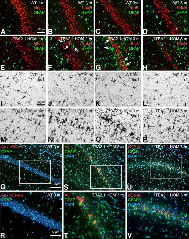
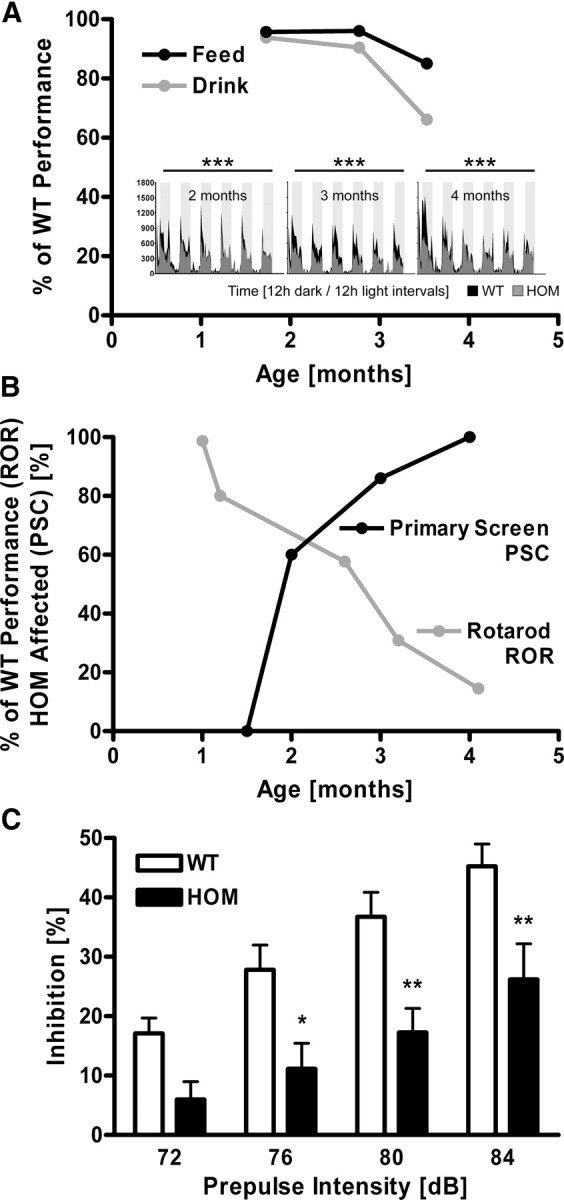
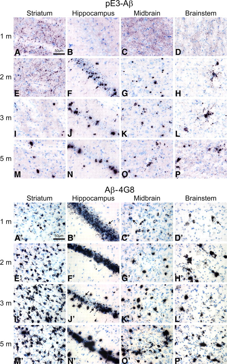
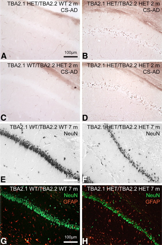
Posttranslational amyloid-β (Aβ) modification is considered to play an important role in Alzheimer's disease (AD) etiology. An N-terminally modified Aβ species, pyroglutamate-amyloid-β (pE3-Aβ), has been described as a major constituent of Aβ deposits specific to human AD but absent in normal aging. Formed via cyclization of truncated Aβ species by glutaminyl cyclase (QC; QPCT) and/or its isoenzyme (isoQC; QPCTL), pE3-Aβ aggregates rapidly and is known to seed additional Aβ aggregation. To directly investigate pE3-Aβ toxicity in vivo, we generated and characterized transgenic TBA2.1 and TBA2.2 mice, which express truncated mutant human Aβ. Along with a rapidly developing behavioral phenotype, these mice showed progressively accumulating Aβ and pE3-Aβ deposits in brain regions of neuronal loss, impaired long-term potentiation, microglial activation, and astrocytosis. Illustrating a threshold for pE3-Aβ neurotoxicity, this phenotype was not found in heterozygous animals but in homozygous TBA2.1 or double-heterozygous TBA2.1/2.2 animals only. A significant amount of pE3-Aβ formation was shown to be QC-dependent, because crossbreeding of TBA2.1 with QC knock-out, but not isoQC knock-out, mice significantly reduced pE3-Aβ levels. Hence, lowering the rate of QC-dependent posttranslational pE3-Aβ formation can, in turn, lower the amount of neurotoxic Aβ species in AD.


Figures

Figure 1.
Figure 1.
Genetic construct, phenotypic hallmarks, and Aβ/pE3–Aβ levels in TBA2.1 and TBA2.1/2.2 mice. In the construct of TBA2.1 and TBA2.2, Aβ(Q3–42) is fused to pre-pro-TRH for product liberation within the secretory pathway. After prohormone convertase cleavage, the N-terminally truncated Aβ peptide is transported into the trans-Golgi and secretory vesicles, in which the N terminus is available to QC for cyclization (A). Transgene expression levels (B) in both lines are gene-dosage dependent, with TBA2.2 expressing ∼80% of transgene compared with HOM TBA2.1 and double HET TBA2.1/TBA2.2 animals expressing ∼90% of transgene. HOM TBA2.1 mice aged 3 months are severely affected in hanging behavior and righting reflex paradigms (C). Both HOM TBA2.1 (D) and double HET TBA2.1/TBA2.2 (D, inset) mice show significantly slower body weight gain over time compared with WT (interaction, p < 0.001); data were analyzed by two-way repeated-measures ANOVA and represent means ± SEM; n ≥ 9 animals per group. Quantification of protein levels by ELISA (E) reveals differential kinetics of Aβ and pE3–Aβ in TBA2.1, with pE3–Aβ peaking at 4 weeks and decreasing to levels of ∼30 ng/g brain at the age of 7 months, whereas Aβ levels increase continuously over time. Significant loss of pE3–Aβ is seen after genetic deletion of QC activity (F, left), whereas a similar deletion of isoQC enzymatic activity has no effect on pE3–Aβ levels (F, right); values expressed as percentage of TBA2.1 HOM/QC-ko WT or isoQC-ko WT, respectively; *p = 0.0101; data were analyzed by one-way ANOVA followed by Newman–Keuls post hoc test and represent means ± SEM; n ≥ 6 animals per group.
Figure 2.
Figure 2.
Neuronal loss and neurodegeneration in TBA2.1 mice. NeuN immunostaining (A–D) shows thinning of the pyramidal cell layer in 3-month-old HOM TBA2.1 mice, most prominent in the medial CA1 area (arrows in C, D) when compared with age-matched WT littermates (A, B). Quantification of neurons in CA1 (E) reveals a significant reduction of neuronal numbers at the age of 3 or 5 months, respectively, in HOM TBA2.1 compared with age-matched WT littermates; ***p < 0.001; data were analyzed by one-way ANOVA followed by Newman–Keuls post hoc test and represent means ± SEM; n = 12 sections per group. Calbindin immunohistochemistry shows reduced immunoreactivity of pyramidal cell somata (PY) and processes in the stratum radiatum (RAD) of HOM TBA2.1 (G, PY and RAD) compared with WT (F). Both the Campbell–Switzer AD pathology stain (CS, H) and the amino–cupric–silver stain for acute neurodegeneration (AmCuAg, I) reveal abundant neuropathological alterations in the medial CA1 of HOM TBA2.1 (H, I) but not in WT littermates (H, I, insets). In semithin sections (J, K), the reduced PY is accompanied by a rarification of dendrites as detected in the RAD of HOM TBA2.1 (K, RAD), which is not observed in WT (J). Dark structures within the PY layer (K, arrow) correspond to severe signs of degeneration. Numerous neurons exhibit a nucleus (n) with an irregular nuclear envelope but the caryoplasm still preserved (L) and abundant cytoplasmic accumulation of dense bodies (L, M, asterisks) as detected by EM (L, M). At a more advanced stage, cell nucleus and cytoplasm are strongly condensed (M).
Figure 3.
Figure 3.
QC and Aβ colocalize in the hippocampal CA1 of HOM TBA2.1 mice at the age of 1 month. Double immunofluorescent labeling of the hippocampal CA1 area with anti-Aβ (red) and anti-QC (green) antibodies (A–F) shows colocalization of enzyme and substrate in the hippocampus of in HOM TBA2.1 animals at 1 month of age (C, F, asterisks). QC immunoreactivity is also detected in scattered interneurons, which do not express Aβ (F, arrow).
Figure 4.
Figure 4.
Progressing astrogliosis, microglial activation, and neuronal loss in the hippocampal CA1 region in TBA2.1 mice is associated with Aβ expression and subsequent pE3–Aβ formation. Double immunofluorescent labeling with anti-NeuN (red) and anti-GFAP (green) antibodies (A–H) shows age-dependent progress of astrogliosis within the CA1 area in HOM animals (E–H) beginning at the age of 2 months (F, arrows) and peaking at 3 months of age (G, arrows), whereas dramatic neuronal loss is visible in 3- and 5-month-old transgenic animals (G, H). Both neuronal loss and astrogliosis are absent in 1-month-old HOM TBA2.1 (E) and in WT animals of all ages (A–D). Immunohistochemical staining with an anti-Iba1 antibody (I–P) shows microgliosis in HOM animals 2 and 3 months of age (N, O, arrows) but not in WT animals of the corresponding ages (J, K), indicating neuroinflammatory processes. Labeling with anti-Aβ/anti-pE3–Aβ (red) and anti-GFAP (green) antibodies and a DAPI nuclear counterstain (blue) (Q–V) shows Aβ and pE3–Aβ immunoreactivity in the field of neuronal loss and gliosis (pyramidal cell layer) at 3 months of age in HOM TBA2.1 (S,T and U,V) but not in WT animals (Q, R), associating pathological processes with the formation of toxic pE3–Aβ from the Aβ transgene.
Figure 5.
Figure 5.
Synaptic dysfunction in TBA2.1 mice. LTP of fEPSP after application of strong tetanus (at time point 0). At 2 months of age, LTP of HOM TBA2.1 does not differ significantly from LTP of age-matched WT littermates (A). At 5 months of age, LTP of HOM TBA2.1 is significantly diminished compared with age-matched WT littermates (C). Analog traces represent typical recordings of single experiments taken 10 min before tetanization (1) and 240 min after tetanization (2). Input–output curve showing the relation between the signal size (fEPSP slope) and the stimulation intensity reveals that, at 2 months of age, HOM TBA2.1 mice show only a slight, nonsignificant reduction of fEPSP amplitude (B). At 5 months of age, the fEPSP amplitude of HOM TBA2.1 is significantly diminished when compared with age-matched WT littermates (D); *p = 0.011, **p = 0.007; data were analyzed by two-way repeated-measures ANOVA (A, C) or by one-way ANOVA (B, D) and represent means ± SEM; n ≥ 12 animals per group.
Figure 6.
Figure 6.
Early onset of behavioral alterations and progressive motor decline in TBA2.1 mice. Behavioral testing in automated home-cage environment reveals decline of free feeding (A, black line) and drinking (A, gray line) over time, as well as significantly altered rearing behavior across 6 d of individual recordings (A, small panels) in HOM TBA2.1 (gray actigrams) compared with age-matched WT littermates (black actigrams); data are expressed as percentage of WT littermate scores (feeding, drinking) or show number of rearing events (small panels) and were analyzed by two-way repeated-measures ANOVA and represent means; ***p < 0.001; n ≥ 7 animals per group. Abnormal performance of HOM TBA2.1 in primary neurobehavioral assessment (B, black line; percentage of animals affected per group) and in the rotarod test (B, gray line; percentage of WT littermate scores) increases over time; n ≥ 7 animals per group. A clear loss of the PPI of the auditory startle reflex in HOM TBA2.1 (C, black bars) compared with age-matched WT littermates (C, open bars) across 72–84 dB prepulse intensity (aged 28 d) is detected; *p < 0.05, **p < 0.01; data were analyzed by two-way repeated-measures ANOVA followed by Bonferroni's post hoc test and represent means ± SEM; n ≥ 14 animals per genotype.
Figure 7.
Figure 7.
Age- and region-dependent accumulation of Aβ and pE3–Aβ in HOM TBA2.1 mice. For a simultaneous front-to-back representation of 25 brains, high-sensitivity immunohistochemistry was performed on MultiBrain sections. Two-dimensional analysis of pE3–Aβ (A–P) and Aβ (A′–P′) immunoreactivity in TBA2.1 mice reveals differential distribution patterns in different brain regions of HOM TBA2.1 mice. Aβ reactivity using the 4G8 anti-Aβ antibody is found both intracellularly in intact cells (e.g., A′–C′, arrows) and extracellularly in evidently disintegrated cells (e.g., J′, arrows). Intracellular Aβ deposits appear to be originating from small, inclusion-like structures (D′, arrow) and seem to be deposited extracellularly after disintegration of the neuron (N′). In contrast, using both a newly generated anti-pE3–Aβ antibody and a commercially available antibody, highest reactivity of pE3–Aβ is detected extracellularly after neuronal disintegration (A–P).
Figure 8.
Figure 8.
Neuropathology in the hippocampal CA1 of TBA2.1/2.2 double transgenic mice. A–D, Campbell–Switzer AD pathology staining of double HETs (B, D) and monoallelic controls (A, C), aged 2 months, reveals CS reactivity in the CA1 pyramidal cell layer of double HET animals only. NeuN immunohistochemistry shows prominent cell loss within the same region in the TBA2.1/TBA2.2 double HET animal (F) but not in the WT control (E, both aged 7 months). Double immunofluorescent labeling of neurons with NeuN (green) and glia with GFAP (red) indicates that neuronal loss is accompanied by gliosis at this time point in double HET (H), but not in WT (G).

Similar articles

Cited by

References

    1. Ashe KH, Zahs KR. Probing the biology of Alzheimer's disease in mice. Neuron. 2010;66:631–645. - PMC - PubMed
    1. Augustin M, Sedlmeier R, Peters T, Huffstadt U, Kochmann E, Simon D, Schöniger M, Garke-Mayerthaler S, Laufs J, Mayhaus M, Franke S, Klose M, Graupner A, Kurzmann M, Zinser C, Wolf A, Voelkel M, Kellner M, Kilian M, Seelig S, Koppius A, Teubner A, Korthaus D, Nehls M, Wattler S. Efficient and fast targeted production of murine models based on ENU mutagenesis. Mamm Genome. 2005;16:405–413. - PubMed
    1. Bayer TA, Wirths O. Review on the APP/PS1KI mouse model: intraneuronal Abeta accumulation triggers axonopathy, neuron loss and working memory impairment. Genes Brain Behav. 2008;7(Suppl 1):6–11. - PubMed
    1. Bayer TA, Wirths O. Intracellular accumulation of amyloid-Beta - a predictor for synaptic dysfunction and neuron loss in Alzheimer's disease. Front Aging Neurosci. 2010;2:8. - PMC - PubMed
    1. Block ML, Zecca L, Hong JS. Microglia-mediated neurotoxicity: uncovering the molecular mechanisms. Nat Rev Neurosci. 2007;8:57–69. - PubMed

Publication types

MeSH terms

Substances

LinkOut - more resources