. 2011 Sep 7;31(36):12927-35.
doi: 10.1523/JNEUROSCI.2301-11.2011.

Fukutin-related protein alters the deposition of laminin in the eye and brain

Affiliations

Fukutin-related protein alters the deposition of laminin in the eye and brain

Mark R Ackroyd et al. J Neurosci. .

Abstract

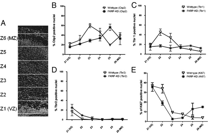
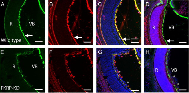
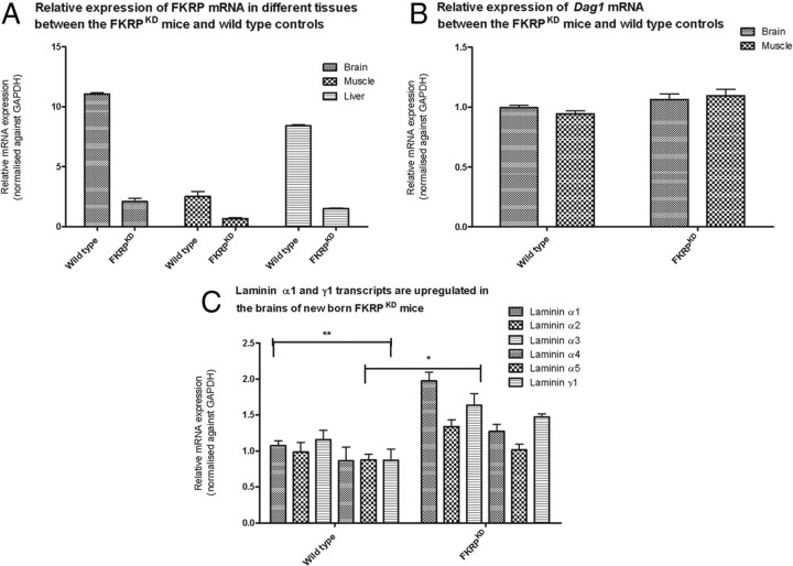
Mutations in fukutin-related protein (FKRP) are responsible for a common group of muscular dystrophies ranging from adult onset limb girdle muscular dystrophies to severe congenital forms with associated structural brain involvement. The defining feature of this group of disorders is the hypoglycosylation of α-dystroglycan and its inability to effectively bind extracellular matrix ligands such as laminin α2. However, α-dystroglycan has the potential to interact with a number of laminin isoforms many of which are basement membrane/tissue specific and developmentally regulated. To further investigate this we evaluated laminin α-chain expression in the cerebral cortex and eye of our FKRP knock-down mouse (FKRP(KD)). These mice showed a marked disturbance in the deposition of laminin α-chains including α1, α2, α4, and α5, although only laminin α1- and γ1-chain mRNA expression was significantly upregulated relative to controls. Moreover, there was a diffuse pattern of laminin deposition below the pial surface which correlated with an abrupt termination of many of the radial glial cells. This along with the pial basement membrane defects, contributed to the abnormal positioning of both early- and late-born neurons. Defects in the inner limiting membrane of the eye were associated with a reduction of laminin α1 demonstrating the involvement of the α-dystroglycan:laminin α1 axis in the disease process. These observations demonstrate for the first time that a reduction in Fkrp influences the ability of tissue-specific forms of α-dystroglycan to direct the deposition of several laminin isoforms in the formation of different basement membranes.


Figures

Figure 1.
Figure 1.
Irregular laminin α-chain deposition in the cerebral cortex of FKRPKD mice. A–H, Coronal sections (20 μm) of the cerebral cortex of newborn wild-type (A–D) and FKRPKD (E–H) mice were stained with a panel of laminin α-chain antibodies. Comparable sections were selected for both the mutants and controls at levels where the left and right hemispheres are juxtaposed and the central hemispheric fissure was clearly present. Laminin α1 immunostaining is mislocalized across the cerebral cortex in the FKRPKD brain (E) compared with wild-type (A) where laminin α1 staining is strictly localized to the pial surface. Laminin α2 and α5 immunostaining is significantly reduced at the pial surface in the FKRPKD brain (F, H) compared with wild-type controls (B, D). Laminin α4 staining is present in the vasculature in both the mutants and controls (C, G) but the staining pattern at the pial surface is highly disorganized in the FKRPKD brain. Scale bars, 50 μm.
Figure 2.
Figure 2.
Radial glial endfeet associate with aberrantly localized laminin α1 in the FKRPKD mice. A–H, Coronal sections (20 μm) of the cerebral cortex of newborn wild-type (A, C, E, G) and FKRPKD mice (B, D, F, H) were immunolabeled with laminin α1 (green) and RC2, a marker for radial glia (red). Nuclei are counterstained with Hoechst 33342 (blue). Laminin α1 immunostaining can be observed throughout the cortex in the FKRPKD cortex (B) compared with wild-type (A) where laminin α1 is restricted to the pial basement membrane. In the wild-type mice the radial glia endfeet form the glial limitans (arrows) and colocalize with laminin α1 at the pial surface. In the FKRPKD the radial glial endfeet do not form a continuous glial limitans at the pial surface and their endfeet can often be observed juxtaposed with the mislocalized laminin α1 (*). C–H represent enhanced regions of A and B, indicated by white boxes. Individual channels for RC2 (E, F) and laminin α1 (G, H) are shown. Scale bars: A, B, 400 μm; C–H, 50 μm.
Figure 3.
Figure 3.
A, Neuronal migration in the cerebral cortex of the FKRPKD mice. a–p, Coronal sections (20 μm) of the cerebral cortex from wild-type (a, d, i–l) and FKRPKD mice (e–h, m–p). Comparable sections were selected in both the mutants and controls at the midline of the cerebral cortex where the left and right hemispheres are juxtaposed. a–h illustrate representative images of cortices costained with Hoechst 33342 (blue), to label the nuclei (a, e); RC2 (red), a marker for radial glia (b, f); and Ctip2 (green), a transcription factor expressed in layer V neurons (c, g). d and h illustrate a merged image of tiles a–c and e–g, respectively. i–l and m–p represent cortices immunolabeled for Tbr-2 (red), a transcription factor expressed in cells of the subventricular zone (j, n) and Ki67 (green), to label proliferating cells. Hoechst 33342 was used to counterstain the nuclei (blue; i, m) and the merged image is shown (l, p). Scale bars, 200 μm. B, Coronal sections (20 μm) of the cerebral cortex from wild-type (a–d) and FKRPKD mice (e–h). Layer VI neurons were labeled with antibodies against Tbr-1 (a, e). Images c and g show immunolabeling for BrdU in the newborn cortex of wild-type and FKRPKD mice following the intraperitoneal injection of BrdU into pregnant females at E15.5. Nuclei were counterstained with Hoechst 33342 (blue) and the merged images are shown in b and f, and d and h. Images were captured at 10× magnification. Scale bars, 200 μm.
Figure 4.
Figure 4.
Distribution of cortical markers and proliferating cells across the cortical plate of FKRPKD and wild-type brains. To quantify the distribution of specific population of neurons across the cortical plate of the FKRPKD mice the cortical plate was arbitrarily divided into 6 equal divisions (A). The distributions of nuclei that were positive for staining with Ctip2 (B), Tbr-1 (C), Tbr-2 (D) and Ki67 (E) are plotted as a percentage of total nuclei.
Figure 5.
Figure 5.
Irregular deposition of laminin α1 at the inner limiting membrane. A–H, Coimmunostaining of wild-type (A–D) and FKRPKD (E–H) for laminin α1 (green; A, E) and laminin 111/211 (red; B, F). The ILM (arrows) is juxtaposed between the retina and vitreous body in the wild-type controls. In the FKRPKD retina this basement membrane is incomplete and ectopic nuclei from the ganglion cell layer (asterisk) of the retina are present in the vitreous body. Images C and G are merged images of A and B, and E and F, respectively. Costaining with RC2 to depict the Müller glia (red) and laminin α1 (green) demonstrate that in the retina of wild-type eye the Müller glia cell endfeet are anchored in the ganglion cell layer, juxtaposed with the ILM (D). In the FKRPKD retina (H) the Müller glia endfeet penetrate through the incomplete basement membrane and are ectopically located in the vitreous body along with ectopically located ganglion cell nuclei. Nuclei are counterstained with Hoechst 33342 (blue). Scale bars, 50 μm.
Figure 6.
Figure 6.
Glycosylated epitopes of α-dystroglycan are expressed at the ILM in a developmental regulated manner. Transverse sections (10 μm) of wild-type mouse eyes at ages E12.5 (A–D) and P0 (E–H) were immunolabeled with a polyclonal pan-laminin antibody (B, F) or the IIH6 antibody (glycosylated α-dystroglycan; C, G). IIH6 staining is clearly present at the ILM of the E12.5 wild-type eye (arrows; C, D) but is not detected in the retina of newborn wild-type mice despite IIH6 immunoreactivity in the sarcolemma of the newborn eye (arrowheads; G, H). Nuclei were visualized with Hoechst 33342 (A, E). Merged images are shown (D, H). Scale bars, 50 μm.
Figure 7.
Figure 7.
Real-time gene expression analysis of Fkrp, Dag1, and laminin in brain and muscle of newborn wild-type and FKRPKD mice. A, Relative expression of FKRP. TaqMan (Applied Biosystems) RT-PCR probes were used to measure relative FKRP mRNA expression in brain, skeletal muscle and liver in the FKRPKD mice compared with age-matched wild-type controls. Expression levels were normalized against endogenous GAPDH mRNA expression. Error bars represent SEM (n = 4). All samples were analyzed as triplicate datasets. B, Relative expression of dystroglycan. TaqMan (Applied Biosystems) RT-PCR probes were used to measure relative Dag1 mRNA expression in brain and skeletal muscle in the FKRPKD mice compared with age-matched wild-type controls. Expression levels were normalized against endogenous GAPDH mRNA expression. Error bars represent SEM (n = 3/4). All samples were analyzed as triplicate datasets. C, Laminin α1 and γ1 mRNA is upregulated in the brain of the FKRPKD mice. TaqMan (Applied Biosystems) RT-PCR probes were used to measure relative laminin α1, α2, α3, α4, α5, and γ1 mRNA expression in the brain of FKRPKD mice compared with age-matched wild-type controls. Expression levels were normalized against endogenous GAPDH mRNA expression. *p < 0.05 (two-tailed t test); **p < 0.005. Error bars represent ± SEM (n = 3). All samples were analyzed as triplicate datasets.

References

    1. Ackroyd MR, Skordis L, Kaluarachchi M, Godwin J, Prior S, Fidanboylu M, Piercy RJ, Muntoni F, Brown SC. Reduced expression of fukutin related protein in mice results in a model for fukutin related protein associated muscular dystrophies. Brain. 2009;132:439–451. - PubMed
    1. Aumailley M, Pesch M, Tunggal L, Gaill F, Fässler R. Altered synthesis of laminin 1 and absence of basement membrane component deposition in (beta)1 integrin-deficient embryoid bodies. J Cell Sci. 2000;113:259–268. - PubMed
    1. Barresi R, Campbell KP. Dystroglycan: from biosynthesis to pathogenesis of human disease. J Cell Sci. 2006;119:199–207. - PubMed
    1. Beggs HE, Schahin-Reed D, Zang K, Goebbels S, Nave KA, Gorski J, Jones KR, Sretavan D, Reichardt LF. FAK deficiency in cells contributing to the basal lamina results in cortical abnormalities resembling congenital muscular dystrophies. Neuron. 2003;40:501–514. - PMC - PubMed
    1. Beltrán-Valero de Bernabé D, Currier S, Steinbrecher A, Celli J, van Beusekom E, van der Zwaag B, Kayserili H, Merlini L, Chitayat D, Dobyns WB, Cormand B, Lehesjoki AE, Cruces J, Voit T, Walsh CA, van Bokhoven H, Brunner HG. Mutations in the O-mannosyltransferase gene POMT1 give rise to the severe neuronal migration disorder Walker-Warburg syndrome. Am J Hum Genet. 2002;71:1033–1043. - PMC - PubMed

Publication types

MeSH terms

LinkOut - more resources