. 2011 Sep 7;3(99):99ra86.
doi: 10.1126/scitranslmed.3002442.

Activation of ERBB2 signaling causes resistance to the EGFR-directed therapeutic antibody cetuximab

Affiliations

Activation of ERBB2 signaling causes resistance to the EGFR-directed therapeutic antibody cetuximab

Kimio Yonesaka et al. Sci Transl Med. .

Abstract

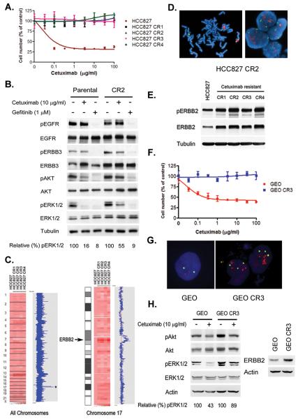
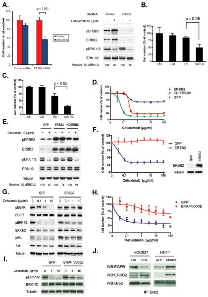
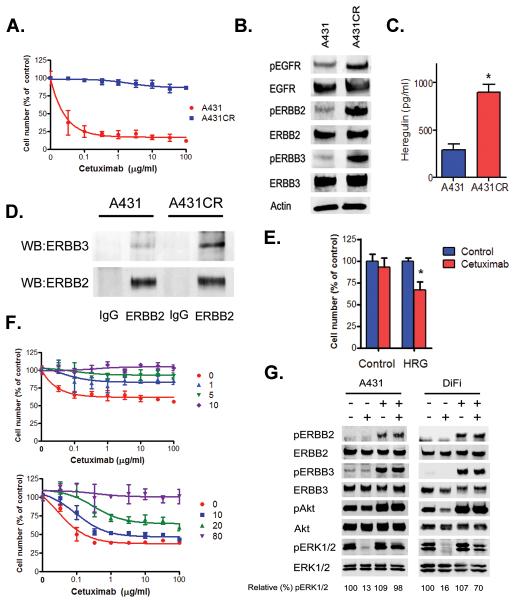
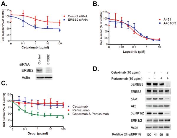
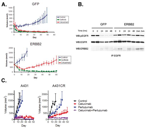
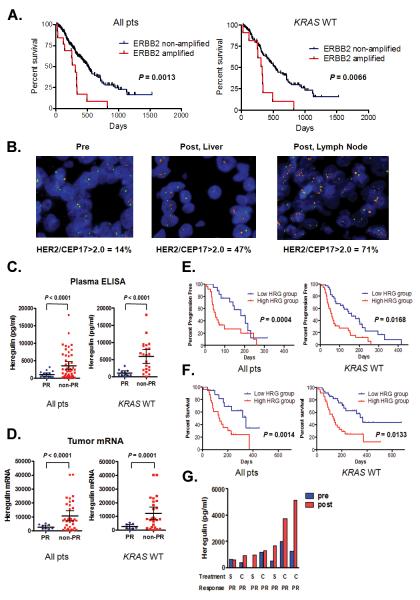
Cetuximab, an antibody directed against the epidermal growth factor receptor, is an effective clinical therapy for patients with colorectal, head and neck, and non-small cell lung cancer, particularly for those with KRAS and BRAF wild-type cancers. Treatment in all patients is limited eventually by the development of acquired resistance, but little is known about the underlying mechanism. Here, we show that activation of ERBB2 signaling in cell lines, either through ERBB2 amplification or through heregulin up-regulation, leads to persistent extracellular signal-regulated kinase 1/2 signaling and consequently to cetuximab resistance. Inhibition of ERBB2 or disruption of ERBB2/ERBB3 heterodimerization restores cetuximab sensitivity in vitro and in vivo. A subset of colorectal cancer patients who exhibit either de novo or acquired resistance to cetuximab-based therapy has ERBB2 amplification or high levels of circulating heregulin. Collectively, these findings identify two distinct resistance mechanisms, both of which promote aberrant ERBB2 signaling, that mediate cetuximab resistance. Moreover, these results suggest that ERBB2 inhibitors, in combination with cetuximab, represent a rational therapeutic strategy that should be assessed in patients with cetuximab-resistant cancers.


Figures

Figure 1
Figure 1. Cetuximab resistant NSCLC and CRC cells maintain ERK 1/2 signaling and contain an ERBB2 amplification
A. Parental and resistant HCC827 CR cells were treated with cetuximab at the indicated concentrations, and viable cells were measured after 72 hours of treatment and plotted (mean +/− SD) relative to untreated controls. B. Parental HCC827 and CR2 cells were treated with cetuximab (10 μg/ml) or gefitinib (1 μM) for 6 hours. Cell extracts were immunoblotted to detect indicated proteins. C. Amplification on chromosome 17 encompassing the ERBB2 locus (asterisk, HCC827 CR cells). The HCC827 CR clones (right) were compared with parental HCC827 cells (first column). The blue curve on the right indicates degree of amplification of each SNP from 0 (left) to 8 (right). Left, genome wide view; right, chromosome 17. D. Metaphase (left) and interphase (right) fluorescence in situ hybridization (FISH) on HCC827 CR2 cells using ERBB2 (red) and CEP 17 (green) probes. The HER2/CEP17 ratio was 4.7. E. Expression of p-ERBB2 and ERBB2 in HCC827 and CR cells. Cell extracts were immunoblotted to detect indicated proteins. F. Parental and resistant GEO CR3 cells were treated with cetuximab at the indicated concentrations, and viable cells were measured after 72 hours of treatment and plotted (mean +/− SD) relative to untreated controls. G. Interphase FISH on GEO and GEO CR3 cells using ERBB2 (red) and CEP 17 (green) probes. HER2/CEP17 ratio ≥ 2 was observed in 50% of GEO CR3 cells. H. (Left) Parental GEO and CR3 cells were treated with cetuximab (10 μg/ml) for 6 hours. Cell extracts were immunoblotted to detect indicated proteins. (Right) Expression of ERBB2 in GEO and GEO CR3 cells.
Figure 2
Figure 2. Inhibition of ERBB2 restores cetuximab sensitivity in cetuximab resistant cancer cell lines
A. Depletion of ERBB2 by an ERBB2 specific shRNA restores sensitivity to cetuximab. Control and ERBB2 shRNA treated HCC827 CR2 cells were treated with cetuximab (10 μg/ml) and viable cells were measured after 72 hours of treatment and plotted relative to untreated controls. Cell extracts were immunoblotted to detect indicated proteins. B. HCC827 CR2 cells were treated with cetuximab (10 μg/ml) or trastuzumab (10 μg/ml) alone or with both agents. Viable cells were measured after 72 hours of treatment and plotted relative to untreated controls. C. GEO CR3 cells were treated with cetuximab (10 μg/ml) or trastuzumab (10 μg/ml) alone or with both agents. Viable cells were measured after 72 hours of treatment and plotted relative to untreated controls. D. HCC827 cells expressing either GFP, ERBB2 or kinase dead (KD) ERBB2 were treated with cetuximab at the indicated concentrations, and viable cells were measured after 72 hours of treatment and plotted (mean +/− SD) relative to untreated controls. E. The indicated cell lines from D. were untreated or treated with cetuximab (10 μg/ml) for 6 hours. Cell extracts were immunoblotted to detect indicated proteins. F. HN11 cells expressing GFP or ERBB2 were treated with cetuximab at the indicated concentrations, and viable cells were measured after 72 hours of treatment and plotted (mean +/− SD) relative to untreated controls. G. HN11 GFP and HN11 ERBB2 cells were treated with indicated concentrations of cetuximab for 6 hours. Cell extracts were immunoblotted to detect indicated proteins. H. HN11 cells expressing GFP or BRAFV600E were treated with cetuximab at the indicated concentrations, and viable cells were measured after 72 hours of treatment and plotted (mean +/− SD) relative to untreated controls. I. Cells from H. were treated with indicated concentrations of cetuximab for 6 hours. Cell extracts were immunoblotted to detect indicated proteins. J. GRB2 co-precipitates with ERBB2 in HCC827 CR2 and HN11 ERBB2 cells. Cell extracts were immunoprecipitated with an anti-Grb2 antibody. The precipitated proteins were determined by immunoblotting with the indicated antibodies.
Figure 3
Figure 3. Heregulin causes resistance to cetuximab
A. Parental and cetuximab resistant A431 cells were treated with cetuximab at the indicated concentrations, and viable cells were measured after 72 hours of treatment and plotted (mean +/− SD) relative to untreated controls. B. A431 CR cells have increased ERBB2 and ERBB3 phosphorylation. Cell extracts were immunoblotted to detect indicated proteins. C. Heregulin in cell culture medium was detected by ELISA from A431 and A431CR cells. *, p = 0.0021; t-test. D. A431 and A431CR cell lysates were immunoprecipitated with anti-ERBB2 antibody. ERBB2 and ERBB3 were detected by immunoblotting. E. Control or HRG siRNAs were transfected into A431CR cells, and cells were treated with 100 μg/ml cetuximab. The percentage of viable cells is shown (mean +/− SD) relative to untreated control. *, p = 0.0007 compared to control; t-test. F. A431 and DiFi cells were treated with cetuximab at the indicated concentrations in the presence of heregulin at the indicated concentrations (ng/ml). Viable cells were measured after 72 hours of treatment and plotted (mean +/− SD) relative to untreated controls. G. A431 and DiFi cells were treated with cetuximab (10 μg/ml) alone, heregulin alone (10 ng/ml for A431; 20 ng/ml for DiFi) or the combination. Cells were lysed, and the indicated proteins were detected by immunoblotting.
Figure 4
Figure 4. ERBB2 inhibition restores cetuximab sensitivity in A431 CR cells
A. Cells transfected with control or ERBB2 siRNA were treated with indicated concentrations of cetuximab. Viable cells were measured after 72 hours of treatment and plotted (mean +/− SD) relative to untreated controls. ERBB2 expression was detected by immunoblotting. B. A431 and A431 CR cells are equally sensitive to lapatinib. C. A431CR cells were treated with cetuximab alone, pertuzumab alone, or a combination of both drugs at the indicated concentrations, and viable cells were measured (mean +/− SD) after 6 days’ treatment. D. A431CR cells were exposed to 10 μg/ml cetuximab alone, 10 μg/ml pertuzumab alone, or a combination of both drugs for 6 h. Cell extracts were immunoblotted to detect the indicated proteins.
Figure 5
Figure 5. Both ERBB2 amplification and heregulin cause cetuximab resistance in vivo
A. Xenografts generated using either HCC827 GFP or ERBB2 cells were treated with vehicle, gefitinib or cetuximab. Vehicle treated mice yielded a median tumor size of 2000 mm3 by 15 days of treatment and were sacrificed. B. Cell extracts from HCC827 GFP or HCC827 ERBB2 tumors treated with cetuximab were immunoprecipitated with anti-EGFR antibody. Precipitated proteins were determined by immunoblotting with the indicated antibodies. C. Xenografts generated using either A431 or A431 CR cells were treated with vehicle, cetuximab alone, pertuzumab alone or the combination of cetuximab and pertuzumab.
Figure 6
Figure 6. Both ERBB2 amplification and heregulin cause drug resistance in cetuximab treated colorectal cancer patients
A. (Left) Overall survival for all CRC patients with (n = 13) and without ERBB2 amplification (n = 220) treated with cetuximab based therapy. Data for KRAS wild type only patients (ERBB2 amplified; n = 11; ERBB2 non-amplified; n = 171). Comparison based on log-rank test. B. ERBB2 FISH from a baseline primary tumor specimen (left) and following acquired cetuximab resistance in two independent drug resistant specimens (right). The patient was initially treated with single agent cetuximab and achieved a PR. ERBB2 (red) and CEP 17 (green). C. Scatter diagram of pre-treatment heregulin concentration in plasma from all (n = 65) or KRAS wild type only (n = 33) CRC patients achieving a PR and those not achieving a PR when treated with cetuximab based therapy. Mean ± 95% CI is shown. D. Scatter diagram of pre-treatment heregulin mRNA expression in tumors from all (n = 44) or KRAS wild type only (n = 34) CRC patients achieving a PR and those not achieving a PR when treated with cetuximab based therapy. Mean ± 95% CI is shown. E. (Left) Progression free survival for all CRC patients treated with cetuximab based therapy divided based on low (n = 35) or high (n = 35) plasma expression. (Right) Data for KRAS wild type only patients (low; n = 18; high n = 24). Comparison based on log-rank test. F. (Left) Overall survival for all CRC patients treated with cetuximab based therapy divided based on low (n = 35) or high (n = 35) plasma expression. (Right) Data for KRAS wild type only patients (low; n = 18; high n = 24). Comparison based on log-rank test. G. Comparisons of plasma levels of heregulin from CRC patients treated with cetuximab based therapy prior to therapy and after the development drug resistance. All patients achieved a PR. S; single agent cetuximab; C; combination with irinotecan.

Comment in

References

    1. Jonker DJ, O’Callaghan CJ, Karapetis CS, Zalcberg JR, Tu D, Au HJ, Berry SR, Krahn M, Price T, Simes RJ, Tebbutt NC, van Hazel G, Wierzbicki R, Langer C, Moore MJ. Cetuximab for the treatment of colorectal cancer. N Engl J Med. 2007;357:2040–2048. - PubMed
    1. Vermorken JB, Mesia R, Rivera F, Remenar E, Kawecki A, Rottey S, Erfan J, Zabolotnyy D, Kienzer HR, Cupissol D, Peyrade F, Benasso M, Vynnychenko I, De Raucourt D, Bokemeyer C, Schueler A, Amellal N, Hitt R. Platinum-based chemotherapy plus cetuximab in head and neck cancer. N Engl J Med. 2008;359:1116–1127. - PubMed
    1. Pirker R, Pereira JR, Szczesna A, von Pawel J, Krzakowski M, Ramlau R, Vynnychenko I, Park K, Yu CT, Ganul V, Roh JK, Bajetta E, O’Byrne K, de Marinis F, Eberhardt W, Goddemeier T, Emig M, Gatzemeier U. Cetuximab plus chemotherapy in patients with advanced non-small-cell lung cancer (FLEX): an open-label randomised phase III trial. Lancet. 2009;373:1525–1531. - PubMed
    1. Kimura H, Sakai K, Arao T, Shimoyama T, Tamura T, Nishio K. Antibody-dependent cellular cytotoxicity of cetuximab against tumor cells with wild-type or mutant epidermal growth factor receptor. Cancer Sci. 2007;98:1275–1280. - PMC - PubMed
    1. Van Cutsem E, Kohne CH, Hitre E, Zaluski J, Chien C. R. Chang, Makhson A, D’Haens G, Pinter T, Lim R, Bodoky G, Roh JK, Folprecht G, Ruff P, Stroh C, Tejpar S, Schlichting M, Nippgen J, Rougier P. Cetuximab and chemotherapy as initial treatment for metastatic colorectal cancer. N Engl J Med. 2009;360:1408–1417. - PubMed

MeSH terms