

Affinity maturation of human CD4 by yeast surface display and crystal structure of a CD4-HLA-DR1 complex
- PMID: 21900604
- PMCID: PMC3179091
- DOI: 10.1073/pnas.1109438108
Affinity maturation of human CD4 by yeast surface display and crystal structure of a CD4-HLA-DR1 complex
Abstract
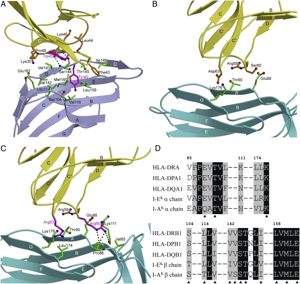
Helper T-cell activation generally requires the coreceptor CD4, which binds MHC class II molecules. A remarkable feature of the CD4-MHC class II interaction is its exceptionally low affinity, which ranges from K(D) = ∼200 μM to >2 mM. Investigating the biological role of the much lower affinity of this interaction than those of other cell-cell recognition molecules will require CD4 mutants with enhanced binding to MHC class II for testing in models of T-cell development. To this end, we used in vitro-directed evolution to increase the affinity of human CD4 for HLA-DR1. A mutant CD4 library was displayed on the surface of yeast and selected using HLA-DR1 tetramers or monomers, resulting in isolation of a CD4 clone containing 11 mutations. Reversion mutagenesis showed that most of the affinity increase derived from just two substitutions, Gln40Tyr and Thr45Trp. A CD4 variant bearing these mutations bound HLA-DR1 with K(D) = 8.8 μM, compared with >400 μM for wild-type CD4. To understand the basis for improved affinity, we determined the structure of this CD4 variant in complex with HLA-DR1 to 2.4 Å resolution. The structure provides an atomic-level description of the CD4-binding site on MHC class II and reveals how CD4 recognizes highly polymorphic HLA-DR, -DP, and -DQ molecules by targeting invariant residues in their α2 and β2 domains. In addition, the CD4 mutants reported here constitute unique tools for probing the influence of CD4 affinity on T-cell activation and development.
Conflict of interest statement
The authors declare no conflict of interest.
Figures


Similar articles
-
Competition of HLA-DR and a beta 2 domain peptide for HIV envelope glycoprotein gp120 binding to CD4.Int Immunol. 1995 Feb;7(2):191-7. doi: 10.1093/intimm/7.2.191. Int Immunol. 1995. PMID: 7734415
-
A recombinant single-chain human class II MHC molecule (HLA-DR1) as a covalently linked heterotrimer of alpha chain, beta chain, and antigenic peptide, with immunogenicity in vitro and reduced affinity for bacterial superantigens.Eur J Immunol. 1997 Aug;27(8):1933-41. doi: 10.1002/eji.1830270817. Eur J Immunol. 1997. PMID: 9295029
-
Disruption of hydrogen bonds between major histocompatibility complex class II and the peptide N-terminus is not sufficient to form a human leukocyte antigen-DM receptive state of major histocompatibility complex class II.PLoS One. 2013 Jul 25;8(7):e69228. doi: 10.1371/journal.pone.0069228. eCollection 2013. PLoS One. 2013. PMID: 23976922 Free PMC article.
-
High-throughput engineering and analysis of peptide binding to class II MHC.Proc Natl Acad Sci U S A. 2010 Jul 27;107(30):13258-63. doi: 10.1073/pnas.1006344107. Epub 2010 Jul 9. Proc Natl Acad Sci U S A. 2010. PMID: 20622157 Free PMC article.
-
Labeling antigen-specific CD4(+) T cells with class II MHC oligomers.J Immunol Methods. 2002 Oct 1;268(1):51-69. doi: 10.1016/s0022-1759(02)00200-4. J Immunol Methods. 2002. PMID: 12213343 Review.
Cited by
-
B cell-reactive triad of B cells, follicular helper and regulatory T cells at homeostasis.Cell Res. 2024 Apr;34(4):295-308. doi: 10.1038/s41422-024-00929-0. Epub 2024 Feb 7. Cell Res. 2024. PMID: 38326478 Free PMC article.
-
The structural basis of αβ T-lineage immune recognition: TCR docking topologies, mechanotransduction, and co-receptor function.Immunol Rev. 2012 Nov;250(1):102-19. doi: 10.1111/j.1600-065X.2012.01161.x. Immunol Rev. 2012. PMID: 23046125 Free PMC article. Review.
-
Comprehensive analysis of MHC class II genes in teleost fish genomes reveals dispensability of the peptide-loading DM system in a large part of vertebrates.BMC Evol Biol. 2013 Nov 26;13:260. doi: 10.1186/1471-2148-13-260. BMC Evol Biol. 2013. PMID: 24279922 Free PMC article.
-
Structural and biophysical insights into the role of CD4 and CD8 in T cell activation.Front Immunol. 2013 Jul 22;4:206. doi: 10.3389/fimmu.2013.00206. eCollection 2013. Front Immunol. 2013. PMID: 23885256 Free PMC article.
-
The versatility of the αβ T-cell antigen receptor.Protein Sci. 2014 Mar;23(3):260-72. doi: 10.1002/pro.2412. Epub 2014 Jan 28. Protein Sci. 2014. PMID: 24375592 Free PMC article. Review.
References
-
- Davis SJ, et al. The nature of molecular recognition by T cells. Nat Immunol. 2003;4:217–224. - PubMed
-
- van der Merwe PA, Davis SJ. Molecular interactions mediating T cell antigen recognition. Annu Rev Immunol. 2003;21:659–684. - PubMed
-
- Cole DK, et al. Human TCR-binding affinity is governed by MHC class restriction. J Immunol. 2007;178:5727–5734. - PubMed
-
- Davis MM, et al. T cells as a self-referential, sensory organ. Annu Rev Immunol. 2007;25:681–695. - PubMed
-
- Janeway CA., Jr The T cell receptor as a multicomponent signalling machine: CD4/CD8 coreceptors and CD45 in T cell activation. Annu Rev Immunol. 1992;10:645–674. - PubMed
Publication types
MeSH terms
Substances
Associated data
- Actions
- Actions
Grants and funding
LinkOut - more resources
Full Text Sources
Other Literature Sources
Molecular Biology Databases
Research Materials
