. 2012 Jan 10;86(1):1-14.
doi: 10.1095/biolreprod.110.089425. Print 2012 Jan.

Immunoprotective properties of primary Sertoli cells in mice: potential functional pathways that confer immune privilege

Affiliations

Immunoprotective properties of primary Sertoli cells in mice: potential functional pathways that confer immune privilege

Timothy J Doyle et al. Biol Reprod. .

Abstract

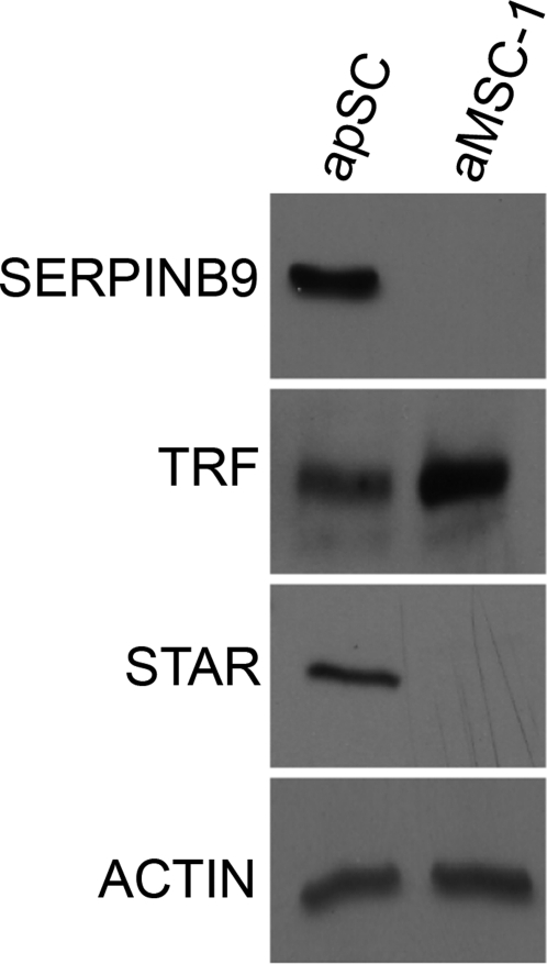
Primary Sertoli cells isolated from mouse testes survive when transplanted across immunological barriers and protect cotransplanted allogeneic and xenogeneic cells from rejection in rodent models. In contrast, the mouse Sertoli cell line (MSC-1) lacks immunoprotective properties associated with primary Sertoli cells. In this study, enriched primary Sertoli cells or MSC-1 cells were transplanted as allografts into the renal subcapsular area of naive BALB/c mice, and their survival in graft sites was compared. While Sertoli cells were detected within the grafts with 100% graft survival throughout the 20-day study, MSC-1 cells were rejected between 11 and 14 days, with 0% graft survival at 20 days posttransplantation. Nonetheless, the mechanism for primary Sertoli cell survival and immunoprotection remains unresolved. To identify immune factors or functional pathways potentially responsible for immune privilege, gene expression profiles of enriched primary Sertoli cells were compared with those of MSC-1 cells. Microarray analysis identified 2369 genes in enriched primary Sertoli cells that were differentially expressed at ±4-fold or higher levels than in MSC-1 cells. Ontological analyses identified multiple immune pathways, which were used to generate a list of 340 immune-related genes. Three functions were identified in primary Sertoli cells as potentially important for establishing immune privilege: suppression of inflammation by specific cytokines and prostanoid molecules, slowing of leukocyte migration by controlled cell junctions and actin polymerization, and inhibition of complement activation and membrane-associated cell lysis. These results increase our understanding of testicular immune privilege and, in the long-term, could lead to improvements in transplantation success.


Figures

FIG. 1.
FIG. 1.
Immunohistochemical analysis of Sertoli cell grafts is shown. Four million apSC were transplanted underneath the kidney capsule of BALB/c mice. Grafts were collected on Days 2 (A, B), 5 (C, D), 14 (E, F), and 20 (G, H) and immunostained for the Sertoli cell marker GATA4 (brown color). Inset shows higher magnification than that in B. The graft is separated from the kidney (K) by a dotted line (A, C, E, G). Sections were counterstained with hematoxylin (blue color). Note that GATA4 also stains kidney epithelial cells; however, grafts and Sertoli cells within the grafts are clearly distinguishable from kidney cells. Arrow, Sertoli cells; I, cellular infiltrate.
FIG. 2.
FIG. 2.
Immunohistochemical analysis of MSC-1 cell grafts is shown. Four million aMSC-1 cells were transplanted underneath the kidney capsule of BALB/c mice. Grafts were collected on Days 1 (A, B), 2 (C, D), 5 (E, F), 11 (G, H), and 20 (I, J) and immunostained for the MSC-1 cell marker large T-antigen (brown color). Inset is a higher magnification of B. The graft is separated from the kidney (K) by a dotted line (A, C, E, G, I). All sections were counterstained with hematoxylin (blue color). Arrow, MSC-1 cells; C, connective tissue; I, cellular infiltrate.
FIG. 3.
FIG. 3.
TUNEL analyses of Sertoli cell and MSC-1 cell grafts are shown. Graft-bearing kidneys were removed 8 days after transplantation of apSC (A, B) or aMSC-1 (C, D), and tissue sections were analyzed for apoptotic cells by TUNEL assay (green color [A, C]). Sections were counterstained with bisBenzimide (Hoechst 33342; blue color [B, D]). A dotted line separates grafts from kidney (K). Insets in A and C show superimposed images of TUNEL-positive cells among Hoechst dye-positive cells at a higher magnification. E) Relative fold changes in the number of apoptotic cells in the SC (grey bar) or MSC-1 cell (black bar) grafts are shown. Data shown are means ± SEM for at least three different experiments per time point. Different letters denote significant differences in means determined by ANOVA followed by the Student t-test method at a P value of ≤0.05.
FIG. 4.
FIG. 4.
Real-time RT-PCR results show normalized signal expression and fold difference between immune-related mRNAs in aMSC1 and those in apSC. The y axis shows the average normalized means ± SEM of the transcript level for aMSC1 (white bar) and apSC (black bar) normalized to those of Rps2 for Cd59, Tgfb1, Il6, Serpinb9, Il11, Csf3, and Serping1 (A), Cd55 and Daf2 (B), and Cd24 (C) transcripts. Real-time RT-PCR was conducted in triplicate using three biological samples per cell type. Relative fold difference between apSC and aMSC1 are designated above each gene. *, significant differences at P ≤ 0.01.
FIG. 5.
FIG. 5.
Real-time RT-PCR results show normalized signal expression and fold difference between known Sertoli cells genes in aMSC1 and those in apSC. The y axis shows the average normalized means ± SEM of transcript level for aMSC1 (white bar) and apSC (black bar) normalized to Rps2 for Lrat, Inha, Fshr, Star, and Clu (A), and Crem (B) transcript levels. Real-time RT-PCR was conducted in triplicate using three biological samples per cell type. Relative fold differences in apSC compared to those in aMSC1 are designated above each gene. *, significant differences at P ≤ 0.01.
FIG. 6.
FIG. 6.
Western blot analysis of SERPINB9, TRF (transferrin), and STAR are shown in apSC and in aMSC-1. Proteins were isolated from apSC and aMSC-1. Equal protein loading was confirmed by immunoblotting the same membranes with an anti-actin antibody. Blots representing three separate experiments are shown.
FIG. 7.
FIG. 7.
Selected immune-related functional pathways are shown. Immune-related functional pathways for cell adhesion and tight junction molecules (A), cytokine–cytokine receptor interactions (B), arachidonic acid and eicosanoid metabolism (C), and classical pathway of the complement cascade (D) are shown. Genes in red are higher and genes in green are lower expression in apSC than in aMSC-1. Boxes touching indicate they are in a complex; black line indicates binding; black arrow indicates direct stimulatory regulation or modification; T indicates inhibitory regulation; thick lines in B represent inner and outer leaflets of membrane.
FIG. 8.
FIG. 8.
A summary diagram is shown. Primary Sertoli cells form tubules with tight junctions (thick red lines) and increase adhesion to lymphocytes, limiting leukocyte migration. A) Under this condition, the Sertoli cell secretory factors (e.g., cytokines and prostanoids) modify the cell-mediated immune response to a type 2 response and suppress inflammation, resulting in immune tolerance and immune privilege. B) Humorally, primary Sertoli cells express complement inhibitors that inhibit activation of the complement cascade and cell lysis, decreasing anaphylotoxin production, leukocyte recruitment, and inflammation and, thus, contribute to immune privilege. Green line, adhesion molecules; green and brown dots, anti-inflammatory molecules secreted by Treg and immature dendritic cells that further promote a type 2 immune response; Y, immunoglobulin.

References

    1. Barker CF, Billingham RE. Immunologically privileged sites. Adv Immunol 1977; 25: 1 54 - PubMed
    1. Whitmore WF, III, Karsh L, Gittes RF. The role of germinal epithelium and spermatogenesis in the privileged survival of intratesticular grafts. J Urol 1985; 134: 782 786 - PubMed
    1. Cameron DF, Whittington K, Schultz RE, Selawry HP. Successful islet/abdominal testis transplantation does not require Leydig cells. Transplantation 1990; 50: 649 653 - PubMed
    1. Selawry HP, Cameron DF. Sertoli cell-enriched fractions in successful islet cell transplantation. Cell Transplant 1993; 2: 123 129 - PubMed
    1. Korbutt GS, Elliott JF, Rajotte RV. Cotransplantation of allogeneic islets with allogeneic testicular cell aggregates allows long-term graft survival without systemic immunosuppression. Diabetes 1997; 46: 317 322 - PubMed

Publication types