. 2011 Sep 8;2(9):e208.
doi: 10.1038/cddis.2011.82.

CYLD regulates keratinocyte differentiation and skin cancer progression in humans

Affiliations

CYLD regulates keratinocyte differentiation and skin cancer progression in humans

J P Alameda et al. Cell Death Dis. .

Abstract

CYLD is a gene mutated in familial cylindromatosis and related diseases, leading to the development of skin appendages tumors. Although the deubiquitinase CYLD is a skin tumor suppressor, its role in skin physiology is unknown. Using skin organotypic cultures as experimental model to mimic human skin, we have found that CYLD acts as a regulator of epidermal differentiation in humans through the JNK signaling pathway. We have determined the requirement of CYLD for the maintenance of epidermal polarity, keratinocyte differentiation and apoptosis. We show that CYLD overexpression increases keratinocyte differentiation while CYLD loss of function impairs epidermal differentiation. In addition, we describe the important role of CYLD in the control of human non-melanoma skin cancer progression. Our results show the reversion of the malignancy of human squamous cell carcinomas that express increased levels of CYLD, while its functional inhibition enhances the aggressiveness of these tumors which progress toward spindle cell carcinomas. We have found that the mechanisms through which CYLD regulates skin cancer progression include the control of tumor differentiation, angiogenesis and cell survival. These findings of the role of CYLD in human skin cancer prognosis make our results relevant from a therapeutic point of view, and open new avenues for exploring novel cancer therapies.


Figures

Figure 1
Figure 1
Properties of transfected HaCaT CYLD cells. (a) Western blot showing CYLD overexpression in one of the H-CYLDWT pools (H-Co, H-Control cells). Tubulin was used as loading control. (b) H-control and H-CYLDWT cells differentiation cultures. Red circles: stratification domes (c) (top) keratin K10 RNA levels in H-control and H-CYLDWT cells detected by northern blotting at the stated times (days); 7S, loading control. (bottom) Graphical representation of the northern blotting signals relative to those of loading controls (arbitrary units). The difference in K10 content between cells of both genotypes is statistically significant at 6, 10 and 13 days of differentiation. (d) Western blot of proteins from H-control, H-CYLDWT and H-CYLDC/S cells in the basal state or after UV stimulation showing an increase in JNK activation in the H-CYLDC/S cells and a reduction in the H-CYLDWT. Analysis of Bax expression. Actin was used as loading control. Western blots were performed 3–4 times
Figure 2
Figure 2
Increased differentiation and apoptosis in H-CYLDWT skin equivalents. (a–d) Skin (6-day) equivalents showing increased differentiation of H-CYLDWT organotypic cultures by histological examination (a and c) and by K1 staining (b and d). Arrow in (a): example of stratified cell; sl, suprabasal layers. (e–h) Skin (12-day) equivalents showing increased apoptosis in the H-CYLDWT skin equivalents by histological observation (e and g) and by anticleaved caspase 3 staining (C3C) (f and h). Arrows in e and g: apoptotic nuclei
Figure 3
Figure 3
Functional analysis of H-CYLDC/S cells. (a) Western blot showing CYLD overexpression in two H-CYLDC/S pools (C/S 1 and C/S 2). (b) Deubiquitination of IKKγ/NEMO (by CYLD) in H-control and H-CYLDC/S cells before (0) or after (30′ and 60′) TNF-α stimulation. IKKγ-immunoprecipitated samples were probed with an antiubiquitin antibody (upper panel) then reprobed with an antibody against IKKγ (lower panel). Observe that TNF-α stimulation failed to increase the polyubiquitination of IKKγ in the H-control cells. By contrast, TNF-α stimulation resulted in significant polyubiquitination of IKKγ in the H-CYLDC/S cells. (c–e) Hematoxylin/eosin staining of 10-day skin equivalents showing the altered morphology of H-CYLDC/S skin equivalents (d and e) versus H-control organotypic cultures (c); arrows in (e and j): foci of invasion. (g) Impaired differentiation of H-CYLDC/S organotypic cultures as studied by involucrin (invol) staining compared with the differentiated H-control skin equivalents (f). (i–j) Cleaved caspase 3 (C3C) immunostaining showing the resistance to apoptosis of the H-CYLDC/S skin equivalents compared with H-control organotypic cultures (h). (k and l) β-catenin expression in H-control, H-CYLDWT and H-CYLDC/S keratinocytes growing as monolayers (l) or in the skin equivalents (k)
Figure 4
Figure 4
Characterization of A-CYLDWT and A-CYLDC/S cells. (a) Western blot analysis of CYLD and Bax expression in total protein extracts from A431 cells of the three genotypes. (b) Monolayer culture of A-control, A-CYLDWT and A-CYLDC/S cells. Red circles: foci of spontaneous differentiation. Note the scattered phenotype of A-CYLDC/S cells and its loose growth in culture. (c) Immunofluorescence showing expression of K10 in A-CYLDWT cells. (d) Western blot of protein extracts from A-control, A-CYLDWT and A-CYLDC/S cells in unstimulated or after UV stimulation. Note the increase in JNK activation in A-CYLDC/S cells and the reduction in the A-CYLDWT cells
Figure 5
Figure 5
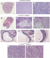
Hematoxilin/eosin staining of A-control, A-CYLDWT and CYLDC/S tumors. (a–c) A-control tumors are solid undifferentiated SCCs. (d–f) A-CYLDWT tumors contain large cavities as a result of tumor degeneration (arrow in d) (g–i); often they also form cystic, differentiated structures. (j–k) A-CYLDC/S SCCs are spindle-shape tumors. Compare the cords of fibroblasts-like cells forming the mutant tumors with the keratinocytes of the A-control SCCs (c). Tumors (10–12) of each type were analyzed
Figure 6
Figure 6
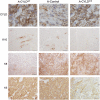
Immunocharacterization of A-control, A-CYLDWT and A-CYLDC/S tumors. Staining with specific antibodies against CYLD, K10, K8 and K5. The increased K8 expression in the A-CYLDC/S tumors, together with lack of K5 and K10 staining are indicative of an aggressive phenotype. Tumors (10–12) of each type were analyzed
Figure 7
Figure 7
CYLDWT expression decreases tumor angiogenesis, while loss of CYLD function enhances the vascularization of skin tumors. (a–c) CD31 staining of blood vessels. Note the smaller size of the vessels in the A-CYLDWT tumors compared with that of A-CYLDC/S SCCs. (d–f) SMA staining. Representative tumors are shown in each panel. Similar data were obtained in five tumors of each genotype. Arrows in (e) indicate the blood vessels. Arrowheads in (f) mark the extremes of the blood vessels. Six tumors of each type were analyzed

References

    1. Fuchs E. Skin stem cells: rising to the surface. J Cell Biol. 2008;180:273–284. - PMC - PubMed
    1. Gazel A, Banno T, Walsh R, Blumenberg M. Inhibition of JNK promotes differentiation of epidermal keratinocytes. J Biol Chem. 2006;281:20530–20541. - PubMed
    1. Weston CR, Wong A, Hall JP, Goad ME, Flavell RA, Davis RJ. The c-Jun NH2-terminal kinase is essential for epidermal growth factor expression during epidermal morphogenesis. Proc Natl Acad Sci USA. 2004;101:14114–14119. - PMC - PubMed
    1. Wheelock MJ, Jensen PJ. Regulation of keratinocyte intercellular junction organization and epidermal morphogenesis by E-cadherin. J Cell Biol. 1992;117:415–425. - PMC - PubMed
    1. Gumbiner B, Stevenson B, Grimaldi A. The role of the cell adhesion molecule uvomorulin in the formation and maintenance of the epithelial junctional complex. J Cell Biol. 1988;107:1575–1587. - PMC - PubMed

Publication types

MeSH terms

Substances