. 2011 Aug;7(8):e1002210.
doi: 10.1371/journal.ppat.1002210. Epub 2011 Aug 25.

A communal bacterial adhesin anchors biofilm and bystander cells to surfaces

Affiliations

A communal bacterial adhesin anchors biofilm and bystander cells to surfaces

Cedric Absalon et al. PLoS Pathog. 2011 Aug.

Abstract

While the exopolysaccharide component of the biofilm matrix has been intensively studied, much less is known about matrix-associated proteins. To better understand the role of these proteins, we undertook a proteomic analysis of the V. cholerae biofilm matrix. Here we show that the two matrix-associated proteins, Bap1 and RbmA, perform distinct roles in the biofilm matrix. RbmA strengthens intercellular attachments. In contrast, Bap1 is concentrated on surfaces where it serves to anchor the biofilm and recruit cells not yet committed to the sessile lifestyle. This is the first example of a biofilm-derived, communally synthesized conditioning film that stabilizes the association of multilayer biofilms with a surface and facilitates recruitment of planktonic bystanders to the substratum. These studies define a novel paradigm for spatial and functional differentiation of proteins in the biofilm matrix and provide evidence for bacterial cooperation in maintenance and expansion of the multilayer biofilm.


Conflict of interest statement

The authors have declared that no competing interests exist.

Figures
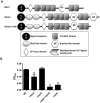
Figure 1
Figure 1. Bap1 and RbmC perform redundant functions in biofilm formation.
(A) Domain analysis of Bap1 and RbmC. Bap1 consists of a signal sequence, an EF hand domain, and a β-prism lectin domain surrounded by six FG-GAP domains. RbmC has two additional StcE-like domains at the N-terminus and an additional β-prism domain at the C-terminus. (B) Quantification of biofilms formed by wild-type V. cholerae (WT), a ΔvpsL mutant, a Δbap1 mutant, a ΔrbmC mutant, and a Δbap1ΔrbmC mutant. * indicate values that are statistically significantly different from wild-type.
Figure 2
Figure 2. Secreted proteins identified in proteomic analyses are retained in the biofilm matrix.
(A) Western blot of cell pellet and supernatant fractions for wild-type V. cholerae with an empty vector or a vector encoding a FLAG-tagged protein as labeled. (B) Transverse sections at the level of the substratum through biofilms containing FLAG-tagged proteins as noted. Proteins were visualized by immunofluorescent staining.
Figure 3
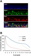
Figure 3. Vertical distribution of Bap1 and RbmA in a native V. cholerae biofilm.
Immunofluorescent imaging of the vertical distribution of (A) Bap1-FLAG and (B) RbmA-FLAG in a native V. cholerae biofilm. Strains harbored a FLAG tag fused to the protein of interest at its chromosomal location. Bacterial DNA was stained with DAPI. (C) Quantification of the fluorescent intensity reflecting Bap1 and RbmA abundance as a function of distance from the substratum.
Figure 4
Figure 4. Vertical distribution of constitutively expressed Bap1 and RbmA in the V. cholerae biofilm.
(A) Vertical section through a biofilm made by co-culturing wild-type V. cholerae carrying either a plasmid encoding Bap1-FLAG or a plasmid encoding RbmA-6X-His. Bacterial DNA was stained with DAPI, and Bap1-FLAG and RbmA-6X-His were visualized with FLAG specific and 6X-His specific antibodies, respectively. (B) Quantification of the total fluorescence due to DAPI, Bap1-FLAG or RbmA-6X-His as a function of biofilm height. Fluorescence in each section was normalized to the maximum fluorescence intensity for that biofilm. Vertical sections and quantifications of fluorescence are representative of the three experimental replicates that were performed in parallel.
Figure 5
Figure 5. Transverse distribution of constitutively expressed Bap1 and RbmA in the V. cholerae biofilm.
A transverse section at the level of the substratum of a biofilm made by co-culture of a Δbap1 mutant carrying a plasmid encoding Bap1-FLAG and wild-type V. cholerae carrying a plasmid encoding RbmA-6X-His. Bacterial DNA was stained with DAPI, and Bap1-FLAG and RbmA-6X-His were visualized by immunofluorescence. Horizontal sections are representative of the three experimental replicates that were performed in parallel. White arrows denote foci of Bap1 staining.
Figure 6
Figure 6. Purified RbmA-FLAG rescues a ΔrbmA mutant.
(A) Quantification of biofilms formed by wild-type V. cholerae or a ΔrbmA mutant carrying either an empty vector (pCTL) or a vector encoding a wild-type rbmA allele (prbmA). The means and standard deviations were calculated from three experimental replicates. While the biofilms formed by the ΔrbmA (pCTL) and the ΔrbmA (prbmA) strains were not significantly different from that formed by wild-type V. cholerae, the ΔrbmA(prbmA) biofilm was significantly greater than the ΔrbmA(pCTL) biofilm (p = 0.004). (B) SDS-PAGE analysis of purified RbmA-FLAG. Protein was visualized with Imperial stain. (C) Biofilms formed by a ΔrbmA mutant rescued with increasing amounts of purified RbmA-FLAG. Biofilms have been vortexed to illustrate fragmentation of the ΔrbmA mutant biofilm.
Figure 7
Figure 7. The Δbap1ΔrbmC mutant biofilm is loosely adherent to the substratum.
Pellicles formed by wild-type V. cholerae, a ΔvpsL mutant, a ΔrbmA mutant, a Δbap1ΔrbmC mutant, a Δbap1ΔrbmC mutant rescued with Bap1-FLAG or RbmA-FLAG expressed from a plasmid, and a Δbap1ΔrbmC mutant co-cultured with a ΔvpsL mutant. Pellicles were photographed without agitation (B), after gentle shaking (S), or after vortexing (V).
Figure 8
Figure 8. Purified Bap1-FLAG restores biofilm formation to a Δbap1ΔrbmC mutant.
(A) SDS-PAGE of affinity purified Bap1-FLAG. A single band is seen at the predicted size of 76 kDA. Protein was visualized with Imperial stain. (B) Quantification of biofilms formed by wild-type V. cholerae (WT), a Δ bap1ΔrbmC mutant rescued with either a control plasmid (pCTL) or a plasmid expressing Bap1-FLAG, and Δ bap1ΔrbmC mutant rescued with purified Bap1 in an 18 nM final concentration. Average measurements and standard deviations were calculated from the results of three experimental replicates. * indicates values that are statistically significantly different from wild-type. (C) Quantification of biofilms made by a Δ bap1ΔrbmC mutant in the presence of increasing amounts of purified Bap1-FLAG. Average measurements and standard deviations were calculated from the results of three experimental replicates. * indicates values that are statistically significantly different from a biofilm formed by a Δbap1ΔrbmC mutant in the absence of purified Bap1. (D) Western analysis of cell extracts prepared from a wild-type biofilm (WT), a biofilm formed with a strain that expresses RbmA-FLAG from the native chromosomal location, and a biofilm formed with a strain that expresses Bap1-FLAG from the native chromosomal location. Blots were probed with an anti-FLAG antibody. Below, the α-subunit of V. cholerae RNA polymerase was visualized with an antibody to the E. coli protein for use as a loading control. Densitometry showed that approximately 16 times less Bap1 is made in biofilm cells as compared with RbmA.
Figure 9
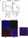
Figure 9. Bap1 is a shared resource; VPS exopolysaccharide is not.
(A) Ratio of biofilm-associated CFU for a panel of co-cultured strains including wild-type V. cholerae (WT), a ΔvpsL mutant, and a Δ bap1ΔrbmC mutant. Strains were labeled by inactivation of the lacZ gene. Label-swapping experiments are shown on right. Black labels indicate a lacZ strain, while blue labels indicate a lacZ + strain. Three experimental replicates are shown for each condition. * indicates ratios that are statistically significantly different from that calculated for the competition of lacZ + wild-type V. cholerae against lacZ wild-type V. cholerae. (B) Transverse section at the level of the substratum through a biofilm formed by co-culture of a Δbap1ΔrbmC mutant with a ΔvpsL mutant carrying chromosomally encoded GFP and a plasmid encoding Bap1-FLAG. The biofilm is comprised primarily of Δbap1ΔrbmC mutant cells surrounded by Bap1-FLAG donated by the ΔvpsL mutant. ΔvpsL mutants are excluded from the biofilm.
Figure 10
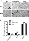
Figure 10. Purified Bap1-FLAG can mediate surface adhesion in the absence of VPS.
(A) Surface adhesion by wild-type V. cholerae, a Δ bap1ΔrbmC mutant, or a ΔvpsL mutant in monolayer minimal medium either alone (no protein), supplemented with purified Bap1-FLAG protein (Bap1), or supplemented with BSA. Phase contrast microscopy was used to obtain images. (B) Surface area coverage of monolayers illustrated in (A). Average measurements and standard deviations were calculated from six microscope fields derived from three experimental replicates.

References

    1. Karatan E, Watnick P. Signals, regulatory networks, and materials that build and break bacterial biofilms. Microbiol Mol Biol Rev. 2009;73:310–347. - PMC - PubMed
    1. Moorthy S, Watnick PI. Genetic evidence that the Vibrio cholerae monolayer is a distinct stage in biofilm development. Mol Microbiol. 2004;52:573–587. - PMC - PubMed
    1. Moorthy S, Watnick PI. Identification of novel stage-specific genetic requirements through whole genome transcription profiling of Vibrio cholerae biofilm development. Mol Microbiol. 2005;57:1623–1635. - PMC - PubMed
    1. Flemming HC, Wingender J. The biofilm matrix. Nat Rev Microbiol. 8:623–633. - PubMed
    1. Kierek K, Watnick PI. Environmental Determinants of Vibrio cholerae Biofilm Development. Appl Environ Microbiol. 2003;69:5079–5088. - PMC - PubMed

Publication types

MeSH terms

Substances