. 2011 Aug;7(8):e1002240.
doi: 10.1371/journal.ppat.1002240. Epub 2011 Aug 25.

Tumor cell marker PVRL4 (nectin 4) is an epithelial cell receptor for measles virus

Affiliations

Tumor cell marker PVRL4 (nectin 4) is an epithelial cell receptor for measles virus

Ryan S Noyce et al. PLoS Pathog. 2011 Aug.

Abstract

Vaccine and laboratory adapted strains of measles virus can use CD46 as a receptor to infect many human cell lines. However, wild type isolates of measles virus cannot use CD46, and they infect activated lymphocytes, dendritic cells, and macrophages via the receptor CD150/SLAM. Wild type virus can also infect epithelial cells of the respiratory tract through an unidentified receptor. We demonstrate that wild type measles virus infects primary airway epithelial cells grown in fetal calf serum and many adenocarcinoma cell lines of the lung, breast, and colon. Transfection of non-infectable adenocarcinoma cell lines with an expression vector encoding CD150/SLAM rendered them susceptible to measles virus, indicating that they were virus replication competent, but lacked a receptor for virus attachment and entry. Microarray analysis of susceptible versus non-susceptible cell lines was performed, and comparison of membrane protein gene transcripts produced a list of 11 candidate receptors. Of these, only the human tumor cell marker PVRL4 (Nectin 4) rendered cells amenable to measles virus infections. Flow cytometry confirmed that PVRL4 is highly expressed on the surfaces of susceptible lung, breast, and colon adenocarcinoma cell lines. Measles virus preferentially infected adenocarcinoma cell lines from the apical surface, although basolateral infection was observed with reduced kinetics. Confocal immune fluorescence microscopy and surface biotinylation experiments revealed that PVRL4 was expressed on both the apical and basolateral surfaces of these cell lines. Antibodies and siRNA directed against PVRL4 were able to block measles virus infections in MCF7 and NCI-H358 cancer cells. A virus binding assay indicated that PVRL4 was a bona fide receptor that supported virus attachment to the host cell. Several strains of measles virus were also shown to use PVRL4 as a receptor. Measles virus infection reduced PVRL4 surface expression in MCF7 cells, a property that is characteristic of receptor-associated viral infections.


Conflict of interest statement

The authors have declared that no competing interests exist.

Figures
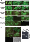
Figure 1
Figure 1. A new receptor for MV is present on smooth airway epithelial cells (SAEC).
(A) Human SAEC were incubated with receptor neutralizing antibodies against CD46 (M75 and B97) or CD150 (IPO-3 and A12) and challenged with the Edmonston vaccine, CD150 Blind, or IC323 wild type strains of MV. Each virus strain contained the EGFP reporter gene. In virus control experiments antibodies against CD46 inhibited infection by Edmonston MV in HeLa cells while antibodies against CD150 blocked infection of Vero-CD150/SLAM by wild type IC323 MV. (B) Marmoset SAEC contain a deletion of the SCR1 domain of CD46 and do not express CD150/SLAM. The panel on the right shows a diagnostic PCR spanning the SCR1 domain revealed by agarose gel electorphoresis in the presence of ethidium bromide, that confirms the deletion in marmoset SAEC. However, the marmoset SAEC could be infected with either the Edmonston or IC323 strains of MV. Virus containing H protein that was mutated in either its CD150 binding site (CD150 Blind) or its CD46 binding site (CD46 Blind) also replicated in the marmoset SAEC. Scale bar = 100 µm.
Figure 2
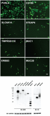
Figure 2. PVRL4 (Nectin 4) can function as an entry factor for IC323-EGFP wtMV.
COS-1 cells were transfected with expression plasmids containing the coding sequences for candidate membrane protein receptors. After 36 hrs the cells were infected with IC323-EGFP wtMV. Virus specific fluorescence was observed between 24–48 hrs infection at 100x magnification using a Leica inverted microscope. Both PVRL4 (Nectin 4) and the positive control CD150/SLAM were capable of converting the non-susceptible COS-1 cells to a virus susceptible phenotype that produced syncytia. Other candidate receptor proteins including SLC6A14, STEAP4, TMPRSS11E, MUC1, ERBB3, and MUC20 were ineffective in producing infections, and yielded only isolated background single-cell infections that did not produce syncytia. Whole cell protein lysates were separated by SDS-PAGE followed by Western Immunoblot using Flag (IB: DDK) and V5 (IB: V5) antibodies to detect expression of these candidate receptors. GAPDH was used as a loading control. Scale bar = 100 µm. See also Figures S2 and S3 in Text S1.
Figure 3
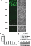
Figure 3. Nectins closely related to PVRL4 cannot function as receptors for wtMV.
COS-1 cells were transfected with expression vectors encoding DDK-tagged versions of PVR, PVRL1, PVRL2, PVRL3, and PVRL4. Control cells were transfected with empty plasmid. After 36 hrs, the transfected cells were infected with IC323-EGFP wtMV and incubated a further 48 hrs. (A) Cells were viewed by fluorescence and phase contrast microscopy. Scale bar  = 200 µm. (B) Virus released from the infected cells was quantified by plaque assay. Data are expressed as the mean of three independent experiments, with error bars showing the SEM. (C) Total cell expression of the transfected proteins was evaluated by Western immunoblots using antibodies directed against the DDK(Flag). (D) Viral proteins were synthesized in PVRL4 transfected cells following MV infection as shown by Western immunoblot using an antibody specific for the viral matrix (M) protein.
Figure 4
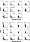
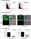
Figure 4. Flow cytometry analysis reveals PVRL4 (Nectin 4) surface expression on cells susceptible for wild type MV infections.
(A) Susceptible cell lines were incubated with a phycoerythrin-conjugated mouse monoclonal antibody that was specific for human PVRL4 (red histogram) or a PE-conjugated mouse IgG2a control antibody (shaded histogram). Cells were washed and analyzed with a Beckman-Coulter ADP Cyan flow cytometer. The Y-axis represents cell counts and the X-axis represents fluorescence intensity. (B) Non-susceptible cell lines were analyzed as described for Panel A.
Figure 5
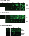
Figure 5. MV infects polarized adenocarinoma cells via either the apical or basolateral surfaces.
Wild type IC323 MV infects (A) MCF7 (breast), (B) NCI-H358 (lung) adenocarcinoma and (C) CHO-PVRL4 cell lines via the apical and basolateral surface in Transwell filter assays. Cells were cultivated in Transwell permeable filter supports at a density of 7.0×10⁵ cells per Transwell filter (24 mm diameter) for 4 days (MCF7 & NCI-H358) or 2 days (CHO-PVRL4). Cells were then infected from either the apical or basolateral side with IC323-EGFP wtMV. At various times post infection fluorescent images were captured. Scale bar = 500 µm.
Figure 6
Figure 6. PVRL4 is localized to both the apical and basolateral surfaces in MCF7 and NCI-H358 cancer cells.
(A) Breast (MCF7 and MDA-MB-231) and (B) lung (NCI-H358 and A549) cancer cell lines were grown to confluence on glass coverslips and then fixed with paraformaldehye, permeabilzed, and stained with goat-anti human PVRL4 antibodies (yellow). Nuclei were visualized with TO-PRO-3 nuclear stain (cyan). Images were captured on a Zeiss upright confocal microscope and analyzed using Zen 2008 image capture software (Zeiss). Scale bar  = 20 µm. (C) Z-sections of MCF7 and NCI-H358 cells stained with PVRL4 (yellow) and TO-PRO-3 (cyan). PVRL4 is localized to both the apical [A] and basolateral [B] surfaces of these cells. White arrowheads indicate the apical expression of PVRL4. (D) Surface biotinylation of MCF7 cells. MCF7 cells were grown for 96 h on transwell filters (24 mm diameter). The cells were incubated with NHS-biotin from either the apical (lanes A) or basolateral (lanes B) side. After lysis, surface proteins were immunoprecipitated with Neutravidin, and immunocomplexes were subjected to SDS-PAGE and Western blot for PVRL4. Glyceraldehyde 3-phosphate (GAPDH) was used as a loading control.
Figure 7
Figure 7. siRNA specific for human PVRL4 inhibits wtMV infections.
MCF7 and NCI-H358 cells were transfected with a scrambled oligonucleotide control (ctrl siRNA) or a siRNA pool specific for PVRL4 (PVRL4 siRNA). The transfected cells were incubated with IC323-EGFP wtMV and images were captured 48 hr post infection. (A) PVRL4 surface expression was detected with a phycoerythrin conjugated PVRL4 antibody following gene knockdown with control siRNA (red line) or PVRL4 siRNA (blue line). (B) PVRL4 siRNA-treated MCF7 and NCI-H358 cells showed less GFP expression compared to ctrl siRNA-treated cells. (C) PVRL4 knockdown results in a decrease in wtMV titres in MCF7 and NCI-H358 cells. Forty-eight hours post infection, cells were harvested and TCID50 virus titrations were performed on Vero-SLAM cells. Data are the means from three independent experiments, and error bars represent the SEM. Scale bar = 100 µm.
Figure 8
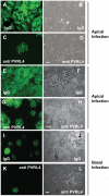
Figure 8. Antibodies specific for human PVRL4 inhibit wtMV infection in MCF7 cells.
MCF7 cells grown on glass coverslips were incubated with 10 µg/ml goat IgG (A,B) or goat anti-PVRL4 (C,D) for 30 min prior to, and during 1 hr adsorption with IC323-EGFP MV via the apical surface. Fluorescence and syncytia formation due to viral infection at 48 hrs was inhibited by the PVRL4 antibody treatment. To determine whether PVRL4 antibodies would also inhibit MV infections via the basolateral route, MCF7 cells were grown on Transwell permeable filter supports as described in Figure 5. Cells were incubated with 25 µg/ml goat IgG on the apical (E,F,G,H) or basal (I,J,K,L) surface with antibodies specific for human PVRL4 or non-immune antibodies (IgG) for 30 min. Cells were subsequently inoculated with IC323-EGFP MV (MOI = 10) for 4 hrs, also in the presence of antibody. Infections were allowed to proceed for 72 hrs and cells were viewed by fluorescence and bright field microscopy. The interaction of goat polyclonal antibodies with PVRL4 blocked MV infection of MCF7 cells via either the apical or basal routes. Scale bar  = 100 µm.
Figure 9
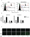
Figure 9. IC323 wtMV binds to cells that stably express human PVRL4.
CHO or CHO stably expressing human PVRL4 (CHO-huPVRL4) were incubated with either 10 or 25 PFU/cell of IC323-EGFP wtMV in the presence of isotype (gIgG Ab) or blocking antibodies against PVRL4 (gPVRL4 Ab) for 1.5 h. Cells were incubated with a MV anti-H primary antibody followed by an anti-mouse alexa fluor 488 conjugated secondary antibody to detect MV-bound cells. (A) Binding of IC323 wtMV to cells stably expressing PVRL4 was detected by FACS. CHO and CHO-huPVRL4 cells were inoculated with MV in the presence of blocking antibody against PVRL4 (gPVRL4, red line) or an isotype control (gIgG, blue line), washed, and incubated with anti-MV hemagglutinin antibody or an isotype matched control antibody (green line). Cells incubated in the absence of virus (Mock, filled histogram) were stained with anti-MV hemagglutinin antibody. Bound MV-specific primary antibody was detected with alexa fluor 488-conjugated goat anti-mouse secondary antibody. The relative fluorescence intensity was measured on a Cyan ADP Flow Cytometer. Inset: Receptor expression was detected with a PE-conjugated PVRL4 antibody (red histogram) or isotype control (filled histogram). (B) Quantification of MV binding to CHO cells expressing huPVRL4 in the presence of blocking antibody to PVRL4 (gPVRL4 Ab). The perecentage of MV-bound cells compared to mock cells was determined using FCS express (De Novo software). Data are expressed as the mean from three independent experiments, with error bars showing the SEM. (C) Infection of CHO and CHO-huPVRL4 cells with varying multiplicities of infection using IC323-EGFP wtMV. Images were captured 48 h post infection. Scale bar  = 500 µm.
Figure 10
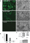
Figure 10. Mouse PVRL4 functions less efficiently as a MV receptor than the human homologue.
COS-1 cells were transfected with expression vectors encoding DDK-tagged human and mouse homologues of PVRL4. Control cells were transfected with empty plasmid. After 36 hrs, the transfected cells were infected with IC323-EGFP wtMV and incubated a further 48 hrs. (A) Cells were viewed by fluorescence and phase contrast microscopy. Scale bar  = 200 µm. (B) Virus released from the infected cells was quantified by plaque assay. Data are expressed as the mean from four independent experiments, with error bars showing the SEM. (C) Total and cell surface expression was evaluated by Western immunoblots using antibodies directed against the DDK(Flag) tag or PVRL4. Surface expression was evaluated following biotinylation of plasma membrane proteins. (D) Viral proteins were synthesized in PVRL4 transfect cells following MV infection as shown by Western immunoblot using an antibody specific for the viral matrix (M) protein.
Figure 11
Figure 11. Surface PVRL4 expression is down regulated following wtMV infection.
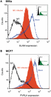
(A) activated marmoset B-cell line B95a or (B) MCF7 cells were infected with IC323-EGFP wtMV. The fusion inhibitory peptide (FIP) was added after the initial virus infection to prevent syncytia formation. At 48 h post-infection SLAM and PVRL4 surface expression was analyzed by FACS. Blue lines, mock-infected cells stained with alexa anti-SLAM antibody (A) or anti-PVRL4 antibody (B); black lines, mock infected cells stained with the anti-mouse IgG2B isotype control antibody; filled orange histogram, cells infected with IC323-EGFP wtMV (MOI = 10) and stained with anti-SLAM (A) or anti-PVRL4 (B) antibodies, respectively. Alexa fluor conjugated 647 secondary antibodies were used to detect SLAM and PVRL4 surface expression. Insets, level of eGFP positive cells following a 48 h infection with IC323-EGFP wtMV. The filled green histogram represents wtMV-infected cells; black lines represent mock-infected cells.

Similar articles

Cited by

References

    1. Enders JF, Katz SL, Holloway A. Development of attenuated measles-virus vaccines. A summary of recentinvestigation. Am J Dis Child. 1962;103:335–340. - PubMed
    1. Moss WJ. Measles control and the prospect of eradication. Curr Top Microbiol Immunol. 2009;330:173–189. - PMC - PubMed
    1. Blake FG, Trask JD. Studies on Measles : I. Susceptibility of Monkeys to the Virus of Measles. J Exp Med. 1921;33:385–412. - PMC - PubMed
    1. de Swart RL. Measles studies in the macaque model. Curr Top Microbiol Immunol. 2009;330:55–72. - PMC - PubMed
    1. Kobune F, Takahashi H, Terao K, Ohkawa T, Ami Y, et al. Nonhuman primate models of measles. Lab Anim Sci. 1996;46:315–320. - PubMed

Publication types

MeSH terms