. 2011 Aug;7(8):e1002238.
doi: 10.1371/journal.pgen.1002238. Epub 2011 Aug 25.

Regulation of Caenorhabditis elegans p53/CEP-1-dependent germ cell apoptosis by Ras/MAPK signaling

Affiliations

Regulation of Caenorhabditis elegans p53/CEP-1-dependent germ cell apoptosis by Ras/MAPK signaling

Rachael Rutkowski et al. PLoS Genet. 2011 Aug.

Abstract

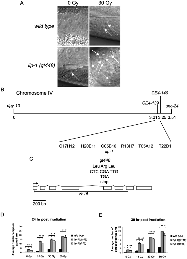
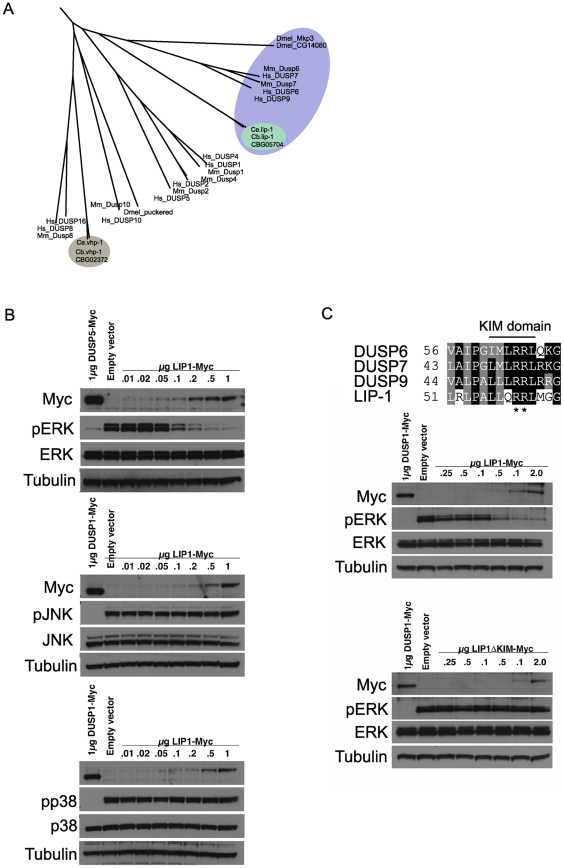
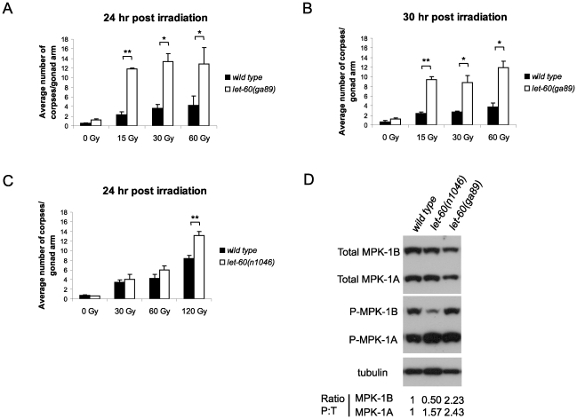
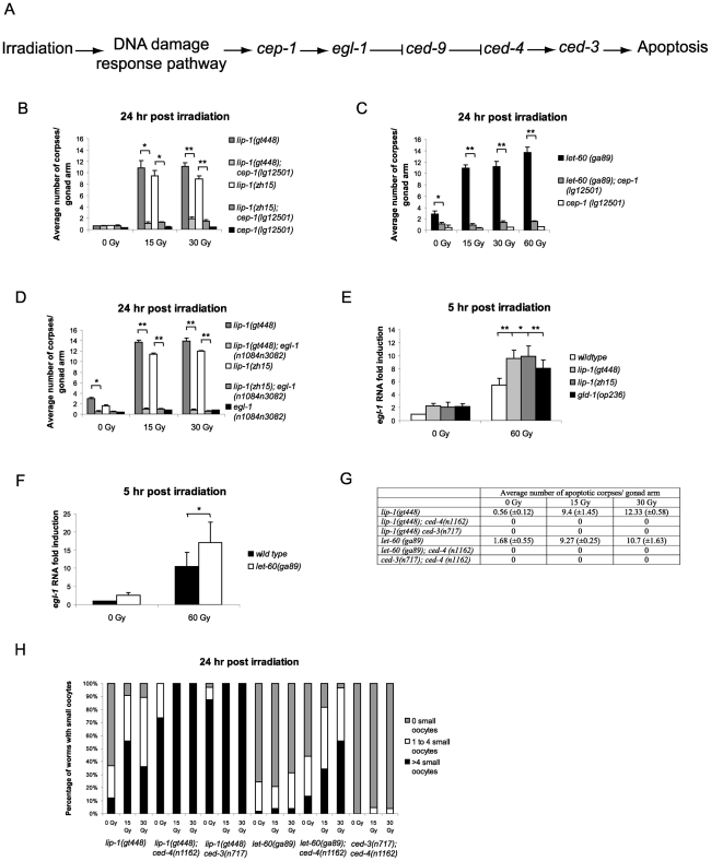
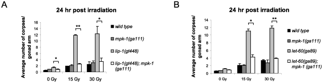
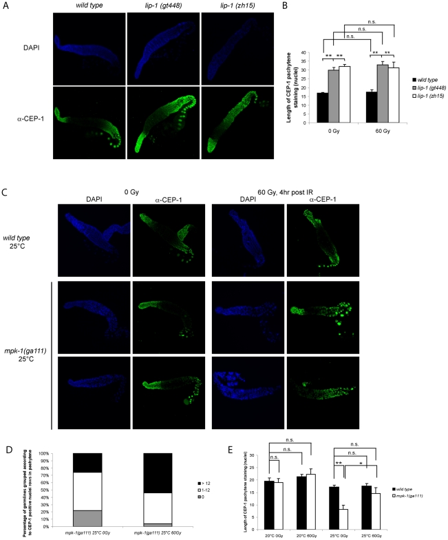
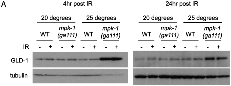
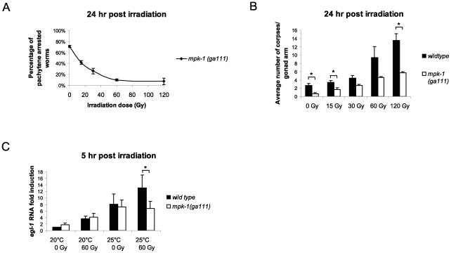
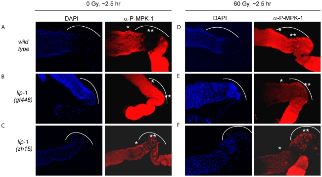
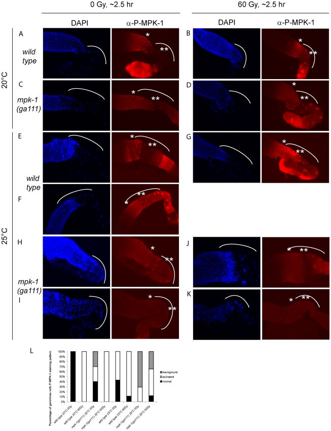
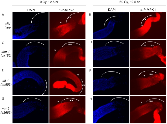
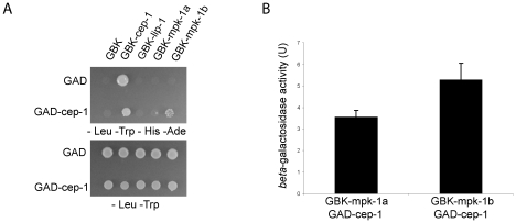
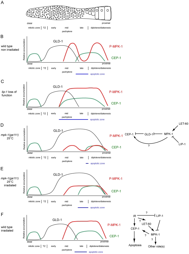
Maintaining genome stability in the germline is thought to be an evolutionarily ancient role of the p53 family. The sole Caenorhabditis elegans p53 family member CEP-1 is required for apoptosis induction in meiotic, late-stage pachytene germ cells in response to DNA damage and meiotic recombination failure. In an unbiased genetic screen for negative regulators of CEP-1, we found that increased activation of the C. elegans ERK orthologue MPK-1, resulting from either loss of the lip-1 phosphatase or activation of let-60 Ras, results in enhanced cep-1-dependent DNA damage induced apoptosis. We further show that MPK-1 is required for DNA damage-induced germ cell apoptosis. We provide evidence that MPK-1 signaling regulates the apoptotic competency of germ cells by restricting CEP-1 protein expression to cells in late pachytene. Restricting CEP-1 expression to cells in late pachytene is thought to ensure that apoptosis doesn't occur in earlier-stage cells where meiotic recombination occurs. MPK-1 signaling regulates CEP-1 expression in part by regulating the levels of GLD-1, a translational repressor of CEP-1, but also via a GLD-1-independent mechanism. In addition, we show that MPK-1 is phosphorylated and activated upon ionising radiation (IR) in late pachytene germ cells and that MPK-1-dependent CEP-1 activation may be in part direct, as these two proteins interact in a yeast two-hybrid assay. In summary, we report our novel finding that MAP kinase signaling controls CEP-1-dependent apoptosis by several different pathways that converge on CEP-1. Since apoptosis is also restricted to pachytene stage cells in mammalian germlines, analogous mechanisms regulating p53 family members are likely to be conserved throughout evolution.


Conflict of interest statement

The authors have declared that no competing interests exist.

Figures

Figure 1
Figure 1. gt448 is an allele of lip-1.
(A) gt448 shows enhanced apoptosis following 30 Gy of ionising irradiation (IR). Wild type (N2) and gt448 L4 larval stage hermaphrodites were irradiated and observed by Nomarski optics 24 hours later. Apoptotic corpses are indicated by arrows. (B) gt448 was mapped to chromosome IV between the visible markers dpy-13 and unc-24. Fine mapping using CB4856 positioned gt488 between the two SNPs CE4-139 and CE4-140. Of the six cosmids in this region, one (C05B10) contained a likely candidate, lip-1. (C) Sequencing of the lip-1 locus identified a C to T change, which introduces a stop codon at amino acid 170. The zh15 allele is also shown. (D and E) Wild type, lip-1(zh15), and lip-1(gt448) hermaphrodite L4 larval stage worms were gamma irradiated at the specified doses and allowed to recover at 20°C for the specified times before apoptotic corpses in the germline were scored by Nomarski optics. Error bars represent the standard error of the mean and the asterisks the p-value for a paired t-test (* p<0.05, ** p<0.01). Animals for each genotype, dose, and time point were assessed in triplicate, scoring a minimum of 15 germlines for each data point.
Figure 2
Figure 2. lip-1 encodes an ERK–specific phosphatase.
(A) Phylogenic tree showing the evolutionary relationships of human (Hs), mouse (Mm), Drosophila (Dmel), C. elegans (Ce) and C. briggsae (Cb) MAPK phosphatases. LIP-1 is highlighted by the green oval within the DUSP6/7/9 cluster (blue oval). The other C. elegans MAPK phosphatase VHP-1 is highlighted by the grey oval. (B) In vivo dephosphorylation assays. Increasing amounts of Myc-tagged LIP-1 was expressed in Cos-1 cells and the phosphorylation of endogenous ERKs 1 and 2 (pERK), JNK (pJNK), or p38 (pp38) in response to either serum stimulation (ERK) or anisomycin (JNK and p38) was monitored by western blotting. Myc-tagged human DUSP5 and DUSP1 were used as positive controls to inactivate ERK and JNK/p38, respectively. (C) Sequence alignment of human DUSP6, DUSP7, DUSP9, and LIP-1, showing the conserved Kinase Interaction Motif (KIM). Increasing amounts of either Myc-tagged wild type or a mutant in which the conserved arginine residues (indicated by asterisks) of the KIM were mutated to alanine were expressed in Cos-1 cells and the phosphorylation of ERK1/2 was assessed. Tagged human DUSP1 was used as a positive control.
Figure 3
Figure 3. let-60(ga89) mutants show enhanced apoptosis following irradiation.
(A and B) Wild type and let-60(ga89) and (C) wild type and let-60(n1046) worms were irradiated and allowed to recover at 20°C, and apoptotic corpses were scored after the stated time points. The scoring of apoptotic corpses was performed as in Figure 1. (D) Activated (phosphorylated) MPK-1 levels differ between let-60(ga89) and let-60(n1046) worms. Protein was extracted from young adult worms (24 hours post L4 larval stage) and equal amounts were loaded onto SDS-PAGE gels. Activated MPK-1 was detected by an anti-P-ERK antibody, total MPK-1 by an anti-ERK antibody, and α-tubulin was used to control for loading. The ratio of phosphorylated MPK-1 to total MPK-1 (Ratio P∶T, normalised against wild type) is shown for the two isoforms.
Figure 4
Figure 4. Epistasis between lip-1(lf) or let-60(ga89) and members of the cep-1 dependent apoptotic pathway.
(A) Scheme of the cep-1 dependent apoptotic pathway. The enhanced apoptosis following irradiation observed in lip-1(lf) and let-60(ga89) mutants is dependent on cep-1 (B and C) and egl-1 (D). In lip-1(lf) (E) and let-60(ga89) (F) worms egl-1 transcription is enhanced, as measured by quantitative real time PCR. Young adult worms were irradiated with the specified dose and mRNA was isolated 5 hours later. mRNA levels for each sample were normalised to γ-tubulin and the fold induction was calculated relative to the levels in wild type untreated worms. Biological experiments were done in triplicate (lip-1) or quadruplicate (let-60(ga89)). Error bars represent the standard error of the mean and asterisks represent the p-value for a paired t-test (* p<0.05, ** p<0.01). (G) Loss of ced-3 or ced-4 suppresses the apoptosis observed in lip-1(gt448) and let-60(ga89) mutants. (H) The small oocyte phenotype of lip-1(gt448) or let-60(ga89) mutants is enhanced by concomitant loss of ced-3 or ced-4. Worms were scored as containing no small oocytes (0), four or less (1 to 4), or greater than four (>4). The scoring of apoptotic corpses was performed as in Figure 1.
Figure 5
Figure 5. The enhanced apoptosis in lip-1(gt448) and let-60(ga89) mutant worms is due to enhanced MPK-1 activity.
(A and B) The increased apoptosis observed in lip-1(gt448) and let-60(ga89) worms is suppressed by mpk-1(ga111ts) when raised at 20°C (permissive temperature). The scoring of apoptotic corpses was performed as in Figure 1.
Figure 6
Figure 6. CEP-1 germline expression is affected by MPK-1 signaling.
(A and B) CEP-1 protein expression is increased in the germline of lip-1 mutants. (A) Germlines from young adult hermaphrodites were dissected and processed for immunofluorescence using an anti-CEP-1 antibody (green). DNA was visualised by DAPI staining (blue). All germlines are orientated the same way with the distal end to the left and the proximal end to the right. (B) For statistical analysis the length of CEP-1 pachytene staining was quantified in germlines of each genotype treated with either 0 Gy or 60 Gy and dissected 4 hours later. Quantification was performed by scoring the number of rows of germ cell nuclei reaching from the pachytene/diplotene border to the most distal point in pachytene in which immunofluorescence was first discernable. At least five germlines were counted for each genotype/treatment. The error bars represent the standard error of the mean and the asterisks the p-value for an unpaired t-test (* p<0.05, ** p<0.01, ‘n.s.’ stands for not significant). (C, D and E) CEP-1 protein is reduced in the germline of mpk-1(ga111ts) mutants raised at the restrictive temperature of 25°C and is increased upon IR treatment. Worms raised at 20°C or 25°C were treated and germlines were dissected and processed for immunofluorescence as in (A). (D) More mpk-1(ga111ts) germlines at 25°C show CEP-1 expression following IR. The number of germlines with pachytene CEP-1 expression were counted and put into three groups: those with expression in more than 12 rows of nuclei, those with expression in 1 to 12 rows, and those with no expression in pachytene stage cells. CEP-1 expression in more than 12 rows implies wild type expression as 13 rows was observed to be the smallest extent of expression in wild type germlines. (E) Average levels of CEP-1 expression are increased in mpk-1(ga111ts) germlines. At least three germlines were counted for each genotype/treatment and the data are presented as in (B).
Figure 7
Figure 7. GLD-1 levels are affected by MPK-1 signaling.
Figure 8
Figure 8. MPK-1 is activated following irradiation and is required for IR–induced egl-1 transcription.
(A) mpk-1(ga111ts) worms were raised at 25°C, which results in a pachytene arrest phenotype with 70% penetrance. mpk-1(ga111ts) L4 hermaphrodites were irradiated with the specified dose and allowed to recover at 25°C for 24 hours. The percentage of worms displaying a pachytene arrested phenotype is shown. (B) These same worms were also examined for the number of apoptotic corpses present as described in Figure 1. (C) Loss of mpk-1 results in reduced egl-1 transcription induction following IR. Wild type and mpk-1(ga111ts) worms were raised at 20°C (permissive temperature) and 25°C (restrictive temperature) and gamma irradiated with the specified dose as young adults (24 hour post L4 larval stage). mRNA was harvested 5 hours later, and the egl-1 transcription assay was performed with quadruple biological replicates as in Figure 4E.
Figure 9
Figure 9. MPK-1 phosphorylation is induced in late pachytene and early diplotene in wild-type germlines following irradiation.
(A and D) Wild type, (B and E) lip-1(gt448), and (C and F) lip-1(zh15) worms (24 hours post L4 larval stage) were treated with 0 or 60 Gy of gamma irradiation and allowed to recover for two hours. Germlines were then dissected and processed for immunofluorescence using an anti-P-MPK-1 antibody (red). Nuclei were stained with DAPI (blue). All images show the germlines from the mid pachytene to the diakenesis region, the loop region (in which the cells are in late pachytene or early diplotene) is highlighted by the dotted arc, the mid pachytene region is highlighted by * and late pachytene by **. All germlines are orientated the same way: distal to the left and proximal to the right.
Figure 10
Figure 10. MPK-1 phosphorylation is slightly increased by irradiation in mpk-1(ga111ts) mutants raised at 25°C.
(A–D) Wild type (A and B) and mpk-1(ga111ts) (C and D) mutants raised at 20°C were treated with 0 or 60 Gy, and allowed to recover for 2 hours before germlines were dissected and processed as in Figure 9. (E–K) Wild type (E–G) and mpk-1(ga111ts) (H–K) mutants raised at 25°C were treated with 0 and 60 Gy, and allowed to recover for 2 hours before germlines were dissected and processed and presented as in Figure 9. (L) Quantification of the number of germlines with P-MPK-1 staining patterns. ‘normal’ describes a wild type non-irradiated staining pattern, ‘activated’ describes a wild type irradiated staining pattern, and ‘background’ describes very faint or no P-MPK-1 staining. At least four germlines were counted for each genotype and treatment.
Figure 11
Figure 11. The activation of MPK-1 by irradiation is not dependent on the DNA damage response pathway.
(A and B) Wild type, (C and D) atm-1(gk186), (E and F) atl-1(tm853), and (G and H) mrt-2(e2663) worms were treated with 0 or 60 Gy, allowed to recover for 2 hours and germlines were then dissected and processed for immunofluorescence and presented as in Figure 9.
Figure 12
Figure 12. CEP-1 and MPK-1 physically interact.
(A) Diploid yeast strains expressing fused Gal4 activation domain (GAD) or Gal4 binding domain (GBK) proteins as indicated were plated onto selective plates (–Leu –Trp –His –Ade) to test for a physical interaction between the fused proteins. –Leu –Trp plates were used to control for growth of the yeast strains. (B) A positive interaction also results in beta-galactosidase expression in these yeast strains. The value shown is the measured activity minus the activity of the control strain GAD-cep-1 GBK-empty. The error bars represent the standard deviation of three replicates.
Figure 13
Figure 13. CEP-1 regulation by MPK-1 signaling.
(A) Scheme of a hermaphrodite germline. Distal is to the left and proximal oocytes are to the right. (B–F) Schematic drawings depicting the relative accumulation of GLD-1 (black), P-MPK-1 (red), and CEP-1 (green) along the length of the hermaphrodite germline (drawings are aligned with panel A). (B) In wild type unirradiated germlines CEP-1 is expressed in the mitotic zone and in late pachytene and diplotene, GLD-1 is expressed from the transition zone (TZ) to mid pachytene, and MPK-1 is phosphorylated in mid pachytene and in the oocytes. The apoptotic zone (blue) is defined by the expression pattern of CEP-1 in pachytene. (C) In lip-1 mutant germlines MPK-1 remains phosphorylated throughout late pachytene and early diplotene. CEP-1 is expressed earlier in pachytene resulting in a wider apoptotic zone. (D) In mpk-1(ga111ts) worms raised at 25°C phosphorylation of MPK-1 is reduced, CEP-1 expression is also reduced, and GLD-1 levels are high (this study) and persist in the proximal region of the germline . (E). Irradiation of mpk-1(ga111ts) worms raised at 25°C results in (partial) activation of P-MPK-1, rescue of CEP-1 expression, but has no effect on GLD-1 protein levels. The regulation of CEP-1 expression by MPK-1 signaling could be due to the effects MPK-1 has on GLD-1 and/or another mechanism (E, right panel). The pattern of GLD-1 expression is adapted from Jones et al. 1996 and Schumacher et al. 2005 . (F) In wild type irradiated germlines CEP-1 and GLD-1 levels are the same as in unirradiated wild type germlines, but MPK-1 is phosphorylated in the late pachytene. MPK-1 phosphorylation could occur through activation of the upstream signaling pathway or through inhibition of LIP-1. IR-activated MPK-1 may affect CEP-1 directly or it may have another role (F, right panel).

Similar articles

Cited by

References

    1. Rutkowski R, Hofmann K, Gartner A. Phylogeny and function of the invertebrate p53 superfamily. Cold Spring Harb Perspect Biol. 2010;2:a001131. - PMC - PubMed
    1. Belyi VA, Ak P, Markert E, Wang H, Hu W, et al. The origins and evolution of the p53 family of genes. Cold Spring Harb Perspect Biol. 2009;2:a001198. - PMC - PubMed
    1. Dotsch V, Bernassola F, Coutandin D, Candi E, Melino G. p63 and p73, the ancestors of p53. Cold Spring Harb Perspect Biol. 2010;2:a004887. - PMC - PubMed
    1. Suh EK, Yang A, Kettenbach A, Bamberger C, Michaelis AH, et al. p63 protects the female germ line during meiotic arrest. Nature. 2006;444:624–628. - PubMed
    1. Schumacher B, Hofmann K, Boulton S, Gartner A. The C. elegans homolog of the p53 tumor suppressor is required for DNA damage-induced apoptosis. Curr Biol. 2001;11:1722–1727. - PubMed

Publication types

MeSH terms