. 2011;6(8):e23977.
doi: 10.1371/journal.pone.0023977. Epub 2011 Aug 25.

Targeted disruption of TgPhIL1 in Toxoplasma gondii results in altered parasite morphology and fitness

Affiliations

Targeted disruption of TgPhIL1 in Toxoplasma gondii results in altered parasite morphology and fitness

Whittney Dotzler Barkhuff et al. PLoS One. 2011.

Abstract

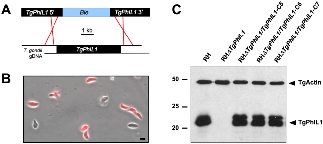
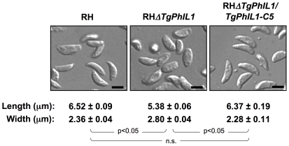
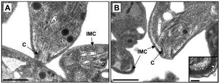
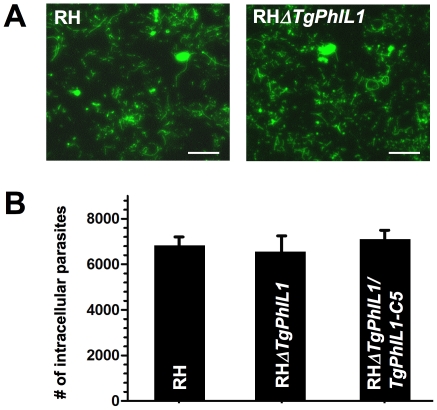
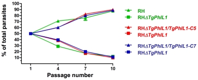
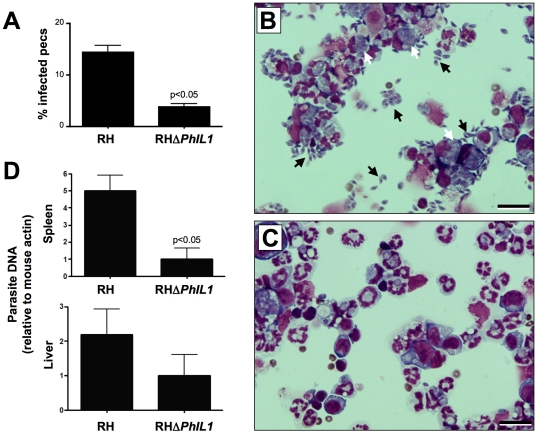
The inner membrane complex (IMC), a series of flattened vesicles at the periphery of apicomplexan parasites, is thought to be important for parasite shape, motility and replication, but few of the IMC proteins that function in these processes have been identified. TgPhIL1, a Toxoplasma gondii protein that was previously identified through photosensitized labeling with 5-[(125)I] iodonapthaline-1-azide, associates with the IMC and/or underlying cytoskeleton and is concentrated at the apical end of the parasite. Orthologs of TgPhIL1 are found in other apicomplexans, but the function of this conserved protein family is unknown. As a first step towards determining the function of TgPhIL1 and its orthologs, we generated a T. gondii parasite line in which the single copy of TgPhIL1 was disrupted by homologous recombination. The TgPhIL1 knockout parasites have a distinctly different morphology than wild-type parasites, and normal shape is restored in the knockout background after complementation with the wild-type allele. The knockout parasites are outcompeted in culture by parasites expressing functional TgPhIL1, and they generate a reduced parasite load in the spleen and liver of infected mice. These findings demonstrate a role for TgPhIL1 in the morphology, growth and fitness of T. gondii tachyzoites.


Conflict of interest statement

Competing Interests: The authors have declared that no competing interests exist.

Figures

Figure 1
Figure 1. Generation of a TgPhIL1 knockout parasite line.
(A) A knockout construct containing a ble cassette, flanked by 3 kb of upstream and downstream noncoding sequence from the TgPhIL1 genomic locus was linearized and transfected into wild-type RH parasites, where it was predicted to integrate into the TgPhIL1 locus by homologous recombination. (B) After three rounds of selection with phleomycin, immunofluorescence with anti-TgPhIL1 antiserum (red) revealed that 6% of the parasites were negative for TgPhIL1, indicating successful disruption of the open reading frame. A combined phase contrast and immunofluorescence image is shown. Scale bar  = 5 µm. (C) Western blotting with anti-TgPhIL1 confirmed the absence of TgPhIL1 expression in the knockout parasites (RHΔTgPhIL1) and restoration of TgPhIL1 expression to approximately wild-type (RH) levels in the complemented clones (RHΔTgPhIL1/PhIL1-C5, C6, C7). The blot was simultaneously probed with anti-actin as a loading control. As seen here, TgPhIL1 sometimes resolves by SDS-PAGE as a tightly spaced doublet, possibly reflecting the presence of an as yet uncharacterized posttranslational modification. Numbers on the left indicate molecular mass in kDa.
Figure 2
Figure 2. Parasites lacking TgPhIL1 are shorter and wider than parasites that express TgPhIL1.
The maximum length and width of individual wild-type (RH), RHΔTgPhIL1, and RHΔTgPhIL1/PhIL1-C5 parasites were measured in differential interference contrast images such as those shown. The experiment was done in triplicate, and numbers indicate the average measurements from 100 parasites/experiment, in µm, plus or minus standard error. The RHΔTgPhIL1 parasites are shorter and wider than either the wild-type or complemented clones (unpaired student's t-test, p<0.05; n.s.  =  not significant). Scale bars  = 5 µm.
Figure 3
Figure 3. Transmission electron microscopy of TgPhIL1 knockout parasites.
The ultrastructure of the conoid (C), IMC, and spacing between the IMC and plasma membrane is indistinguishable in (A) wild-type (RH) and (B) RHΔTgPhIL1 parasites. Scale bars  = 1 µm. Inset shows an enlarged portion of the RHΔTgPhIL1parasite from the lower left corner of panel B; the normal trilaminar structure of the pellicle is evident (arrowheads). Scale bar  = 0.1 µm.
Figure 4
Figure 4. Motility and invasion of the TgPhIL1 knockout parasites.
(A) Motility assays were performed on wild-type (RH) and RHΔTgPhIL1 parasites. The density and shape of the trails deposited by RHΔTgPhIL1 parasites are qualitatively indistinguishable from those deposited by RH parasites. Scale bars  = 10 µm. (B) Laser-scanning cytometry (LSC)-based invasion assays were performed on wild-type (RH), RHΔTgPhIL1, and RHΔTgPhIL1/PhIL1-C5 parasites. Samples were analyzed in duplicate and the results shown are the average from two independent experiments, plus or minus standard error. No significant difference was seen in the ability of the RHΔTgPhIL1 parasites to invade host cells when compared to either RH or RHΔTgPhIL1/TgPhIL1-C5 parasites (unpaired student's t-test, p>0.05).
Figure 5
Figure 5. Growth competition of TgPhIL1 knockout parasites in culture.
Equal numbers of parasites from the different lines were mixed pairwise and serially passaged through monolayers of HFF cells. After every three passages, immunofluorescence was performed and 500 parasites were counted and scored as either positive or negative for TgPhIL1. Each color (red, green, and blue) indicates a pair of parasite lines passaged together. Wild-type (RH) parasites (green triangles) and each of the complemented clones (RHΔTgPhIL1/TgPhIL1-C5 [red triangles] and RHΔTgPhIL1/TgPhIL1-C7 [blue triangles] outgrew the RHΔTgPhIL1 parasites (squares) at all time points (passages 4, 7 and 10). Each experiment was performed in duplicate, and the results shown are the average of the three experiments, plus or minus standard error. The difference between the growth of each of the pairs was significant at all passage numbers >1 (unpaired student's t-test, p<0.05).
Figure 6
Figure 6. Survival and dissemination of TgPhIL1 knockout parasite in a mouse model of infection.
Mice were infected intraperitoneally with 1×10⁴ wild-type (RH) or RHΔTgPhIL1 tachyzoites. (A-C) Peritoneal exudate cells (PECs) were harvested seven days post-infection, spun onto coverslips and examined for the presence of parasites. Panel A shows that PECs from RH-infected mice carry a significantly higher parasite load than those from RHΔTgPhIL1-infected mice (unpaired student t-test, p<0.05). Results shown are the average from five mice, plus or minus standard error. Panels B and C show representative examples of the coverslips from which the data in panel A were derived (Panel B, RH-infected PECs; Panel C, RHΔTgPhIL1-infected PECs). Black arrows indicate extracellular parasites and white arrows intracellular parasites. Scale bars  = 20 µm. (D) DNA was harvested from spleen and liver samples seven days post-infection, and qPCR was performed to determine the amount of parasite DNA present. Results shown are the average parasite loads from five mice, plus or minus standard error. The liver and spleen each contained a higher load of RH than RHΔTgPhIL1 parasites, although only the differences observed in the spleen were statistically significant (unpaired student t-test, p<0.05).

References

    1. Mital J, Ward GE. Current and Emerging Approaches to Studying Invasion in Apicomplexan Parasites. Subcell Biochem. 2007;47:1–32. - PubMed
    1. Kim K, Weiss LM. Toxoplasma gondii: the model apicomplexan. Int J Parasitol. 2004;34:423–432. - PMC - PubMed
    1. Mann T, Beckers C. Characterization of the subpellicular network, a filamentous membrane skeletal component in the parasite Toxoplasma gondii. Mol Biochem Parasitol. 2001;115:257–268. - PubMed
    1. Morrissette NS, Sibley LD. Cytoskeleton of apicomplexan parasites. Microbiol Mol Biol Rev. 2002;66:21–38. - PMC - PubMed
    1. Dubremetz J-F, Torpier G, Maurois P, Prensier G, Sinden R. [Structure of the sporozoite pellicule of Plasmodium yoelii: a cryofracture study]. C. R. Acad Sci Paris. 1979;288:623–626.

Publication types

Substances