. 2011 May;2(5):523-37.
doi: 10.1177/1947601911417177.

The Role of BH3-Only Proteins in Tumor Cell Development, Signaling, and Treatment

Affiliations

The Role of BH3-Only Proteins in Tumor Cell Development, Signaling, and Treatment

Rana Elkholi et al. Genes Cancer. 2011 May.

Abstract

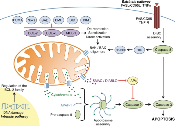
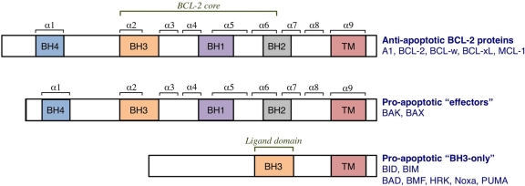
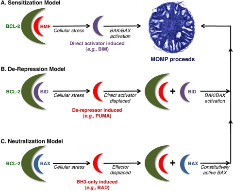
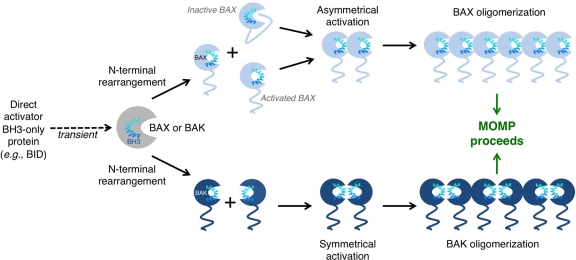
Tumor cells have devised several strategies to block the mitochondrial pathway of apoptosis despite endogenous or pharmacological cues to die. This process of cell death proceeds through the coordinated regulation of multiple anti-apoptotic and pro-apoptotic BCL-2 family proteins that ultimately impinge on the integrity of the outer mitochondrial membrane. Once compromised, mitochondria release pro-apoptotic factors to promote caspase activation and the apoptotic phenotype. Within the BCL-2 family exists a subclass of pro-apoptotic members termed the BH3-only proteins, which directly and/or indirectly functionally regulate the remaining anti- and pro-apoptotic BCL-2 proteins to compromise mitochondria and engage apoptosis. The focus of this review is to discuss the cellular and pharmacological regulation of the BH3-only proteins to gain a better understanding of the signaling pathways and agents that regulate this class of proteins. As the BH3-only proteins increase cellular sensitivity to pro-apoptotic agents such as chemotherapeutics, numerous small-molecule BH3 mimetics have been developed and are currently in various phases of clinical trials. Toward the end of the review, the discovery and application of the small-molecule BH3 mimetics will be discussed.

Keywords: BCL-2 family; BH3-only proteins; apoptosis; cancer; mitochondria.


Conflict of interest statement

The author(s) declared no potential conflicts of interest with respect to the research, authorship, and/or publication of this article.

Figures

Figure 1.
Figure 1.
The regulation of apoptosis by the BCL-2 family. The intrinsic pathway is initiated by cellular stress signals such as DNA damage and growth factor/cytokine withdrawal. These intrinsic signals lead to the transcriptional upregulation and/or activation of pro-apoptotic BH3-only proteins such as BIM, BID, BAD, BMF, Noxa, and PUMA. These proteins are able to bind anti-apoptotic members of the family (e.g., BCL-2, BCL-xL, and/or MCL-1) and inhibit their activity. In addition to binding and inhibiting anti-apoptotic proteins, direct activator BH3-only proteins (e.g., BID and BIM) also bind and activate the effector molecules BAK and BAX. Once activated, BAK and BAX homo-oligomerize and subsequently form pores in the outer mitochondrial membrane, leading to mitochondrial outer membrane permeabilization. Pro-apoptotic proteins such as cytochrome c and SMAC/DIABLO are subsequently released from the intermembrane space into the cytosol. Cytochrome c forms a complex with APAF-1 and pro-caspase 9, whereas SMAC/DIABLO binds the inhibitors of apoptosis proteins (IAPs), which normally bind and inhibit initiator as well as effector caspases. Both steps allow for the dimerization and activation of caspase 9, subsequent cleavage and activation of effector caspase 3, and the apoptotic phenotype. The extrinsic apoptotic pathway is activated by extracellular signals such as death ligands, including FASL/CD95L and tumor necrosis factor (TNF) α. Binding of these ligands to their receptors (FAS and TNF receptor, respectively) causes receptor trimerization and subsequent recruitment of several factors that form the death-inducing signaling complex (DISC). The DISC activates initiator caspase 8, which subsequently activates effector caspase 3. Crosstalk can occur between the extrinsic and intrinsic pathways following the DISC assembly. Activated caspase 8 is able to cleave and activate the pro-apoptotic BH3-only protein BID, leading to C8-BID, which can simultaneously initiate the intrinsic apoptotic pathway through effector molecule activation.
Figure 2.
Figure 2.
The BCL-2 family of proteins. The family is divided into anti- and pro-apoptotic members. The anti-apoptotic members, which include A1, BCL-2, BCL-xL, BCL-w, and MCL-1, share homology in all 4 BCL-2 homology (BH) domains (BH1-4). BCL-2 homology domains 1 to 3 comprise the BCL-2 structural core and create the hydrophobic groove. It is this “BCL-2 groove” that is the binding site for the BH3 domains of pro-apoptotic members. The pro-apoptotic members are subdivided into “effector” proteins and “BH3-only” proteins. The effector proteins also contain homology domains 1 to 4, whereas the BH3-only proteins contain only one BH domain, the BH3, which binds the anti-apoptotic proteins. Many BCL-2 proteins also contain a hydrophobic transmembrane domain (TM).
Figure 3.
Figure 3.
Current models for BH3-only protein functions. (A) Sensitization model. In this scenario, a sensitizer BH3-only protein (e.g., BMF) binds to and inactivates anti-apoptotic members of the family (e.g., BCL-2). Following apoptotic stimulation, a direct activator (e.g., BIM) is induced and subsequently activates the effector proteins (BAK and BAX), which homo-oligomerize and form pores in the outer mitochondrial membrane (OMM), and mitochondrial outer membrane permeabilization (MOMP) proceeds. By inhibiting the anti-apoptotic repertoire, the sensitizer BH3-only protein prevents the inhibition of direct activators, thereby lowering the threshold for effector activation and “sensitizing” the cell to apoptotic stimulation. (B) De-repression model. In the de-repression model, a direct activator is bound to and sequestered by an anti-apoptotic protein. In response to cellular stress, a de-repressor BH3-only protein (e.g., PUMA) is induced and binds the anti-apoptotic protein, thereby displacing the direct activator and allowing it to activate the effectors, and MOMP follows. (C) Neutralization model. This final model postulates that even in the absence of an apoptotic stimulus, a cell contains activated BAK and BAX, but they are bound to and inhibited by the anti-apoptotic proteins. When the cell encounters stress, the BH3-only proteins (e.g., BAD) are induced and bind the anti-apoptotics, releasing the effector proteins. The effectors can then oligomerize and induce MOMP. This model negates the requirement for direct activator-effector interactions.
Figure 4.
Figure 4.
BAK/BAX activation and oligomerization promote mitochondrial outer membrane permeabilization (MOMP). BAK and BAX can be activated through interaction with direct activator BH3-only proteins and the outer mitochondrial membrane. These interactions induce conformational changes that lead to N-terminal rearrangement and the exposure of the BAK/BAX BH3 domain. In the case of BAK, rearrangement leads to the transient exposure of its BH3 domain, which subsequently binds into the hydrophobic groove of another BAK monomer. BAK dimers are able to form multimers through an α6-α6 interaction. High molecular weight species of BAK form pores in the outer mitochondrial membrane (OMM) and induce MOMP. A similar mechanism exists for BAX activation. N-terminal rearrangement of BAX leads to exposure of its BH3 domain, as well as releasing α5, α6, and α9 from the hydrophobic groove. Exposure of these α-helices allows BAX to insert into the OMM. A single BAX molecule can “propagate” this activation signal through binding of its exposed BH3 domain to inactive BAX molecules. Many activated BAX monomers form high molecular weight species once in the OMM to promote MOMP.

Similar articles

Cited by

References

    1. Barr PJ, Tomei LD. Apoptosis and its role in human disease. Biotechnology (N Y). 1994;12:487-93 - PubMed
    1. McDonnell TJ, Korsmeyer SJ. Progression from lymphoid hyperplasia to high-grade malignant lymphoma in mice transgenic for the t(14; 18). Nature. 1991;349:254-6 - PubMed
    1. Strasser A, Whittingham S, Vaux DL, et al. Enforced BCL2 expression in B-lymphoid cells prolongs antibody responses and elicits autoimmune disease. Proc Natl Acad Sci U S A. 1991;88:8661-5 - PMC - PubMed
    1. Thompson CB. Apoptosis in the pathogenesis and treatment of disease. Science. 1995;267:1456-62 - PubMed
    1. Watanabe-Fukunaga R, Brannan CI, Copeland NG, Jenkins NA, Nagata S. Lymphoproliferation disorder in mice explained by defects in Fas antigen that mediates apoptosis. Nature. 1992;356:314-7 - PubMed

LinkOut - more resources