. 2012 Sep;33(9):2147-60.
doi: 10.1002/hbm.21347. Epub 2011 Aug 30.

The role of the anterior cingulate cortex in emotional response inhibition

Affiliations

The role of the anterior cingulate cortex in emotional response inhibition

Jacobo Albert et al. Hum Brain Mapp. 2012 Sep.

Abstract

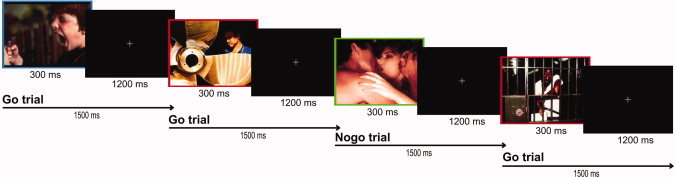
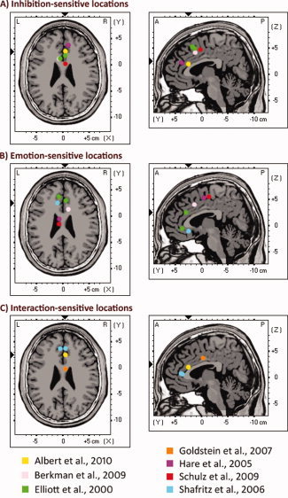
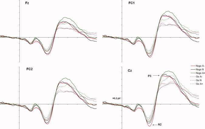
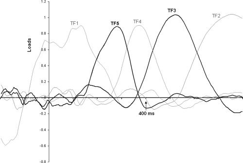
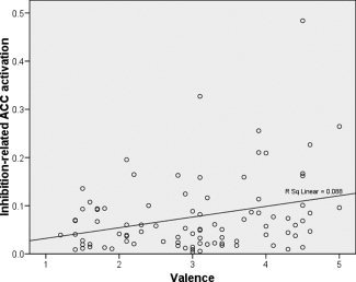
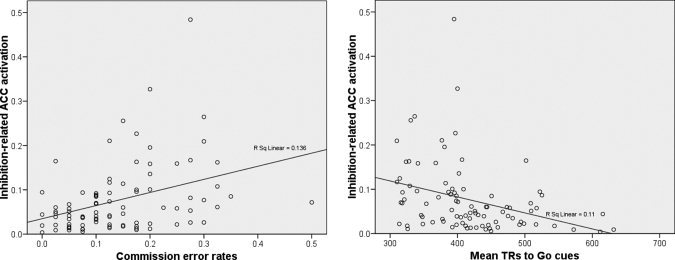
Although the involvement of the anterior cingulate cortex (ACC) in emotional response inhibition is well established, there are several outstanding issues about the nature of this involvement that are not well understood. The present study aimed to examine the precise contribution of the ACC to emotion-modulated response inhibition by capitalizing on fine temporal resolution of the event-related potentials (ERPs) and the recent advances in source localization. To this end, participants (N = 30) performed an indirect affective Go/Nogo task (i.e., unrelated to the emotional content of stimulation) that required the inhibition of a motor response to three types of visual stimuli: arousing negative (A-), neutral (N), and arousing positive (A+). Behavioral data revealed that participants made more commission errors to A+ than to N and A-. Electrophysiological data showed that a specific region of the ACC at the intersection of its dorsal and rostral subdivisions was significantly involved in the interaction between emotional processing and motor inhibition. Specifically, activity reflecting this interaction was observed in the P3 (but not in the N2) time range, and was greater during the inhibition of responses to A+ than to N and A-. Additionally, regression analyses showed that inhibition-related activity within this ACC region was associated with the emotional content of the stimuli (its activity increased as stimulus valence was more positive), and also with behavioral performance (both with reaction times and commission errors). The present results provide additional data for understanding how, when, and where emotion interacts with response inhibition within the ACC.


Figures

Figure 1
Figure 1
Schematic illustration of the emotional Go/NoGo task. An animation reproducing several trials of the affective Go/Nogo task as well as their temporal characteristics can be seen at http://www.uam.es/carretie/grupo/EmoGoNogo.htm. [Color figure can be viewed in the online issue, which is available at wileyonlinelibrary.com.]
Figure 2
Figure 2
Depiction of location of MNI coordinates used to define the regions of interest (ROIs) within the ACC and the functionally related areas of the medial wall. For presentation purposes, coordinates were collapsed on a representative brain slice at Z = 25 (axial view) and Y = 4 (sagittal view). Exact coordinates are given in Table III. These coordinates represent the center of the ROIs (radius = 9 mm). All these locations have been shown to be activated in previous studies on emotional response inhibition. A: ACC locations previously associated with response inhibition. B: ACC locations previously associated with emotional processing. C: ACC locations previously associated with the interaction of emotional processing and response inhibition. An interactive animation reproducing the location of each MNI coordinate employed to define the ROIs (projected one by one on sagittal, axial, and coronal slices of the Colin brain) can be seen at http://www.uam.es/carretie/grupo/cooACC.htm. [Color figure can be viewed in the online issue, which is available at wileyonlinelibrary.com.]
Figure 3
Figure 3
Grand averages at fronto‐central areas, where N2 and P3 are clearly visible. [Color figure can be viewed in the online issue, which is available at wileyonlinelibrary.com.]
Figure 4
Figure 4
tPCA: Factor loadings after promax rotation. Temporal factors 5 (N2) and 3 (P3) are drawn in black.
Figure 5
Figure 5
Scatter plots of valence (1, negative, to 5, positive) and inhibition‐related ACC activation (centroid coordinates of the ROI: 5 25 20) showing the regression line. Number of cases: 90 [30 participants × 3 conditions (NogoA−, NogoN, NogoA+)].
Figure 6
Figure 6
A: Scatter plots of commission error rates and inhibition‐related ACC activation (centroid coordinates of the ROI: 5 25 20) showing the regression line. B: Scatter plots of mean RTs to Go cues and inhibition‐related ACC activation (centroid coordinates of the ROI: 5 25 20) showing the regression line. In both analyses the number of cases was 90 [30 participants × 3 conditions (NogoA−, NogoN, NogoA+)].

References

    1. Albert J, López‐Martín S, Carretié L ( 2010): Emotional context modulates response inhibition: Neural and behavioral data. Neuroimage 49: 914–921. - PubMed
    1. Beckmann M, Johansen‐Berg H, Rushworth MFS ( 2009): Connectivity‐based parcellation of human cingulate cortex and its relation to functional specialization. J Neurosci 29: 1175–1190. - PMC - PubMed
    1. Berkman ET, Burklund L, Lieberman MD ( 2009): Inhibitory spillover: Intentional motor inhibition produces incidental limbic inhibition via right inferior frontal cortex. Neuroimage 47: 705–712. - PMC - PubMed
    1. Beste C, Saft C, Andrich J, Gold R, Falkenstein M ( 2008): Response inhibition in Huntington's disease—A study using ERPs and sLORETA. Neuropsychologia 46: 129. - PubMed
    1. Bishop S, Duncan J, Brett M, Lawrence AD ( 2004): Prefrontal cortical function and anxiety: Controlling attention to threat‐related stimuli. Nat Neurosci 7: 184–188. - PubMed

Publication types