. 2012 Feb;18(3-4):310-9.
doi: 10.1089/ten.TEA.2011.0193. Epub 2011 Oct 19.

Three-dimensional cell grafting enhances the angiogenic efficacy of human umbilical vein endothelial cells

Affiliations

Three-dimensional cell grafting enhances the angiogenic efficacy of human umbilical vein endothelial cells

Suk Ho Bhang et al. Tissue Eng Part A. 2012 Feb.

Abstract

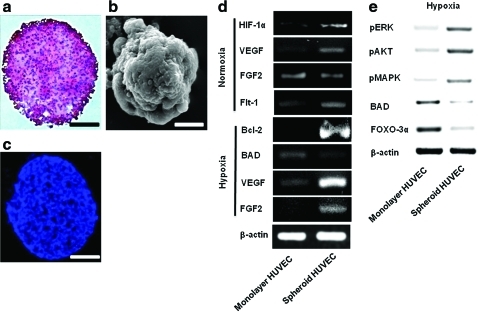
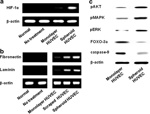
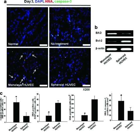
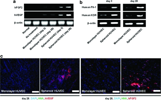
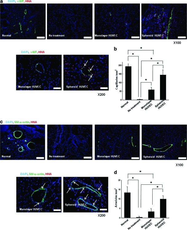
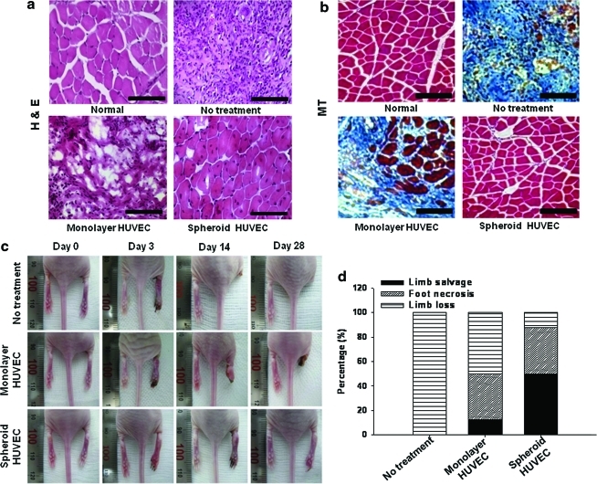
Despite the great potential of cell therapy for ischemic disease, poor cell survival after engraftment in ischemic tissue limits its efficacy. Here we tested a hypothesis that three-dimensionally grafted human umbilical vein endothelial cell (HUVEC) spheroids would exhibit improved angiogenic efficacy following transplantation into mouse ischemic limbs compared with HUVECs prepared by conventional two-dimensional monolayer culture. One day after surgical induction of hindlimb ischemia in athymic mice, HUVECs cultured in monolayer or HUVEC spheroids were transplanted intramuscularly into ischemic limbs. Four weeks after the treatment, in the spheroid HUVEC transplantation group, we observed increased hypoxia-inducible factor-1α expression, decreased apoptosis, and increased HUVEC survival in the ischemic tissue compared with the monolayer HUVEC transplantation group. Transplantation of HUVEC spheroids also resulted in enhanced and prolonged secretion of paracrine factors as well as enhanced expression of factors involved in the recruitment of circulating angiogenic progenitor cells. In summary, transplantation of HUVECs as spheroids enhanced cell survival, increased paracrine factor secretion, and showed a potential as a therapeutic method to treat ischemic tissue damages by promoting angiogenesis.


Figures

FIG. 1.
FIG. 1.
Representative properties of HUVEC spheroids. Formation of HUVEC spheroids in vitro (a–c) and alteration in gene expression by spheroid formation in vitro (d) at 3 days of culture. (a) H&E staining. Scale bar=100 μm. (b) Scanning electron microscopy image. Scale bar=100 μm. (c) TUNEL staining. Scale bar=100 μm. (d) RT-PCR and (e) western blot analysis. HUVEC, human umbilical vein endothelial cell; H&E, hematoxylin and eosin; RT-PCR, reverse transcription–polymerase chain reaction. Color images available online at www.liebertonline.com/tea
FIG. 2.
FIG. 2.
Enhanced expression of cell survival factors and preservation of HUVEC ECMs by transplantation of HUVEC spheroids into ischemic tissues. (a) RT-PCR analysis of human-specific HIF-1α expression in ischemic tissue at 1 day after transplantation of HUVEC spheroids or monolayer HUVECs. (b) RT-PCR analysis to examine human ECM components (fibronectin and laminin) present in ischemic tissues at 1 day after transplantation. (c) Western blot analysis for the cell survival regulators pAKT, pMAPK, and pERK and for the proapoptotic factors FOXO-3α and caspase-9 of monolayer HUVECs and HUVEC spheroids at 3 days after transplantation. HIF-1α, hypoxia-inducible factor-1α; ECM, extracellular matrix; pMAPK, phospho-mitogen activated protein; pERK, phospho-extracellular signal-regulated kinase; pAKT, phospho-AKT.
FIG. 3.
FIG. 3.
Enhanced HUVEC survival in ischemic limb muscle by grafting as spheroids. (a) Immunofluorescent staining for caspase-3 (green) of ischemic limb tissues retrieved at 3 days after HUVEC grafting. HUVECs were double-stained with HNA (red). Apoptosis of transplanted HUVECs (arrows) was reduced in the spheroid group. Scale bars=100 μm. (b) RT-PCR analysis for human proapoptotic (BAD) and antiapoptotic (Bcl-2) factor at 3 days after HUVECs grafting. (c) The ratio of caspase-3-positive cells (apoptotic cells) to HNA-positive cells (transplanted HUVECs) and the ratio of HNA-positive cells (transplanted cells) to 4,6-diamidino-2-phenylindole-positive cells (total cells) in the ischemic region (*p<0.05). HNA-positive cells and HNA/caspase-3 double-positive cells were counted as well. HNA, human nuclear antigen. Color images available online at www.liebertonline.com/tea
FIG. 4.
FIG. 4.
Enhanced and prolonged secretion of angiogenic growth factors from HUVECs in ischemic limb muscle by grafting as spheroids. RT-PCR analysis using human-specific primers for (a) VEGF and FGF2 and (b) angiogenic growth factor activation marker at 3 and 28 days after transplantation. (c) Double-immunofluorescent staining for HNA and the human-specific angiogenic growth factors VEGF and FGF2 (red) of ischemic limb tissues at 28 days after HUVECs (green). Scale bars=200 μm. VEGF, vascular endothelial growth factor; FGF2, fibroblast growth factor 2. Color images available online at www.liebertonline.com/tea
FIG. 5.
FIG. 5.
Enhanced angiogenesis by transplantation of HUVECs as spheroids. Double-immunofluorescent staining for HNA and mouse-specific (a) vWF and (c) SM α-actin of the ischemic hindlimb tissues retrieved at 28 days after HUVEC transplantation. Scale bars indicate 200 μm at 100×and 100 μm at 200×. The quantification of (b) capillary density and (d) arteriole density in the ischemic region (*p<0.05 between groups compared). Quantification of the number of HUVECs incorporated into (e) capillaries and (f) arterioles in the ischemic region (*p<0.05). (g) RT-PCR analysis of human-specific vWF, SM α-actin, HNA, and proliferating cell nucleus antigen of the ischemic hindlimb tissues at 28 days after transplantation. (h) Western blot analysis for ICAM, VCAM, NG2, and MMP-2 in the ischemic hindlimb tissues at 28 days after transplantation. White arrow indicates double stained cells (vWF+/HNA+, or SM α-actin+/HNA+). vWF, von Willebrand factor; SM, smooth muscle; ICAM, intercellular adhesion molecule; VCAM, vascular cell adhesion molecule; MMP, matrix metalloproteinase. Color images available online at www.liebertonline.com/tea
FIG. 5.
FIG. 5.
Enhanced angiogenesis by transplantation of HUVECs as spheroids. Double-immunofluorescent staining for HNA and mouse-specific (a) vWF and (c) SM α-actin of the ischemic hindlimb tissues retrieved at 28 days after HUVEC transplantation. Scale bars indicate 200 μm at 100×and 100 μm at 200×. The quantification of (b) capillary density and (d) arteriole density in the ischemic region (*p<0.05 between groups compared). Quantification of the number of HUVECs incorporated into (e) capillaries and (f) arterioles in the ischemic region (*p<0.05). (g) RT-PCR analysis of human-specific vWF, SM α-actin, HNA, and proliferating cell nucleus antigen of the ischemic hindlimb tissues at 28 days after transplantation. (h) Western blot analysis for ICAM, VCAM, NG2, and MMP-2 in the ischemic hindlimb tissues at 28 days after transplantation. White arrow indicates double stained cells (vWF+/HNA+, or SM α-actin+/HNA+). vWF, von Willebrand factor; SM, smooth muscle; ICAM, intercellular adhesion molecule; VCAM, vascular cell adhesion molecule; MMP, matrix metalloproteinase. Color images available online at www.liebertonline.com/tea
FIG. 6.
FIG. 6.
Histological analysis and physiological status of the hindlimbs at 28 days after the treatment. (a) H&E staining and (b) Masson's trichrome staining. Blue region indicates fibrosis. Scale bars=100 μm. (c) Photographs of mouse hindlimbs at days 0, 3, 14, and 28 after transplantation. Photomicrographs and photographs are representative of each group. (d) Physiological status of the ischemic hindlimbs at 28 days after transplantation. Color images available online at www.liebertonline.com/tea

References

    1. Chekanov V. Akhtar M. Tchekanov G. Dangas G. Shehzad M.Z. Tio F. Adamian M. Colombo A. Roubin G. Leon M.B. Moses J.W. Kipshidze N.N. Transplantation of autologous endothelial cells induces angiogenesis. Pacing Clin Electrophysiol. 2003;26:496. - PubMed
    1. Kim E.J. Li R.K. Weisel R.D. Mickle D.A. Jia Z.Q. Tomita S. Sakai T. Yau T.M. Angiogenesis by endothelial cell transplantation. J Thorac Cardiovasc Surg. 2001;122:963. - PubMed
    1. Calvani M. Rapisarda A. Uranchimeg B. Shoemaker R.H. Melillo G. Hypoxic induction of an HIF-1alpha-dependent bFGF autocrine loop drives angiogenesis in human endothelial cells. Blood. 2006;107:2705. - PMC - PubMed
    1. Zhang M. Methot D. Poppa V. Fujio Y. Walsh K. Murry C.E. Cardiomyocyte grafting for cardiac repair: graft cell death and anti-death strategies. J Mol Cell Cardiol. 2001;33:907. - PubMed
    1. Nakamura Y. Yasuda T. Weisel R.D. Li R.K. Enhanced cell transplantation: preventing apoptosis increases cell survival and ventricular function. Am J Physiol Heart Circ Physiol. 2006;291:H939. - PubMed

Publication types

MeSH terms

Substances

LinkOut - more resources