. 2011 Nov;155(3):349-61.
doi: 10.1111/j.1365-2141.2011.08850.x. Epub 2011 Sep 9.

Novel epitope evoking CD138 antigen-specific cytotoxic T lymphocytes targeting multiple myeloma and other plasma cell disorders

Affiliations

Novel epitope evoking CD138 antigen-specific cytotoxic T lymphocytes targeting multiple myeloma and other plasma cell disorders

Jooeun Bae et al. Br J Haematol. 2011 Nov.

Erratum in

  • Br J Haematol. 2011 Dec;155(5):642

Abstract

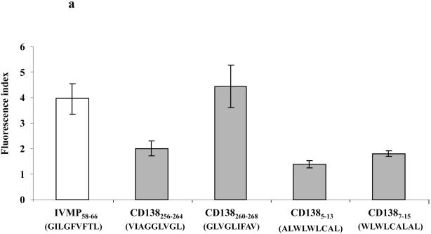
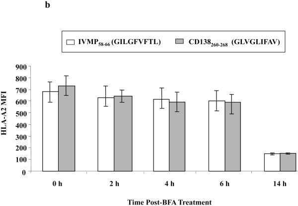
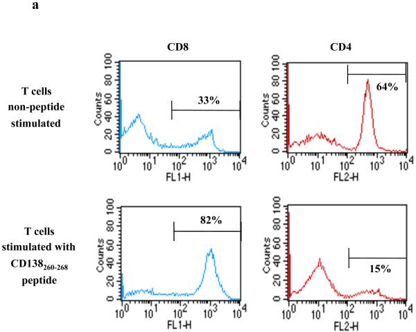
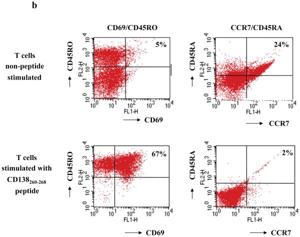
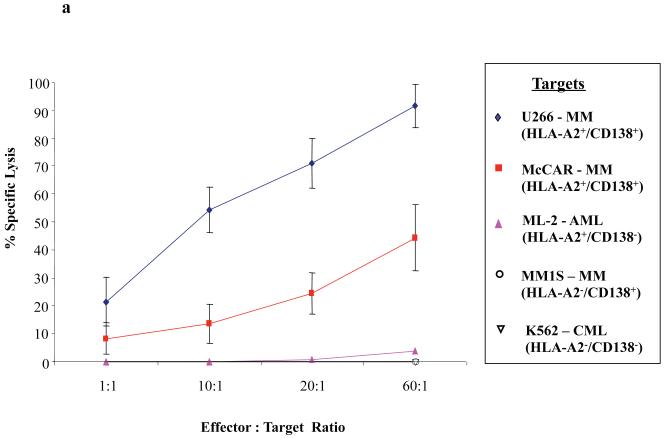
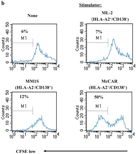
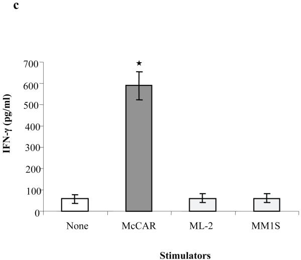
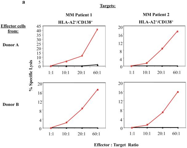
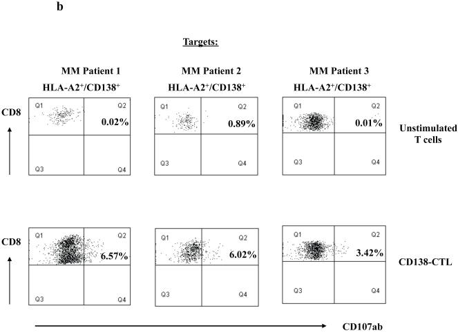
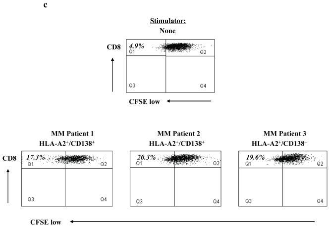
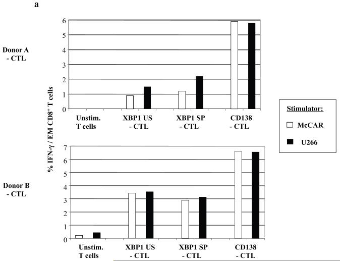
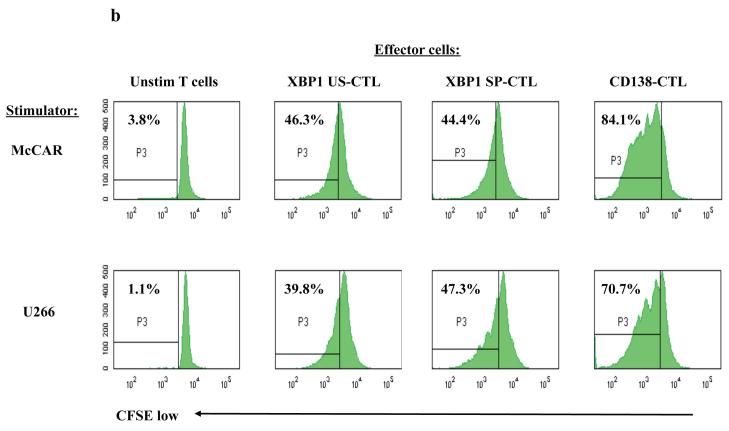
The development of an immunotherapeutic strategy targeting CD138 antigen could potentially represent a new treatment option for multiple myeloma (MM). This study evaluated the immune function of CD138 peptide-specific cytotoxic T lymphocytes (CTL), generated ex vivo using an HLA-A2-specific CD138 epitope against MM cells. A novel immunogenic HLA-A2-specific CD138(260-268) (GLVGLIFAV) peptide was identified from the full-length protein sequence of the CD138 antigen, which induced CTL specific to primary CD138(+) MM cells. The peptide-induced CD138-CTL contained a high percentage of CD8(+) activated/memory T cells with a low percentage of CD4(+) T cell and naive CD8(+) T cell subsets. The CTL displayed HLA-A2-restricted and CD138 antigen-specific cytotoxicity against MM cell lines. In addition, CD138-CTL demonstrated increased degranulation, proliferation and γ-interferon secretion to HLA-A2(+) /CD138(+) myeloma cells, but not HLA-A2(-) /CD138(+) or HLA-A2(+) /CD138(-) cells. The immune functional properties of the CD138-CTL were also demonstrated using primary HLA-A2(+) /CD138(+) cells isolated from myeloma patients. In conclusion, a novel immunogenic CD138(260-268) (GLVGLIFAV) peptide can induce antigen-specific CTL, which might be useful for the treatment of MM patients with peptide-based vaccine or cellular immunotherapy strategies.


Figures

Figure 1
Figure 1. Affinity and stability of CD138260-268 (GLVGLIFAV) peptide binding to HLA-A2 molecules
(a) The HLA-A2 binding affinity of CD138260-268 (GLVGLIFAV) peptide on T2 cells was analysed by flow cytometry. T2 cells were pulsed with CD138 peptide (50 μg/ml) or influenza virus matrix protein58-66 (30 μg/ml) in AIM-V serum-free media. After overnight incubation, the cells were washed to remove unbound peptides and stained with HLA-A2-FITC mAb. The binding affinity of peptides was determined as the Fluorescence Index (FI), which was calculated as [mean channel fluorescence of T2 cells pulsed with the peptide plus β2-microglobulin ÷ mean channel fluorescence of T2 cells pulsed with β2-microglobulin]. Among the peptides tested, the CD138260-268 (GLVGLIFAV) peptide displayed the highest HLA-A2 binding affinity. The values represent the mean ± SE of three separate experiments. (b) The HLA-A2 binding stability of CD138260-268 (GLVGLIFAV) peptide on T2 cells was measured at different time points. After overnight peptide pulsing, the T2 cells were incubated with Brefeldin A, followed by staining with HLA-A2 FITC mAb at 0, 2, 4, 6 or 14 h incubation. The peptide binding stability was measured as HLA-A2 MFI. The stability of CD138260-268 (GLVGLIFAV) peptide was comparable to influenza virus matrix protein58-66 (GILGFVFTL) peptide, which was used as an HLA-A2-specific positive control. The values represent the mean ± SE of three separate experiments.
Figure 2
Figure 2. Generation of activated memory cells in CTL cultures stimulated with CD138260-268 (GLVGLIFAV) peptide
HLA-A2+/CD3+ T cells were stimulated weekly with irradiated antigen-presenting cells pulsed with CD138260-268 peptide (GLVGLIFAV). One week after their fourth stimulation, the phenotype of the T cell cultures was analysed by flow cytometry. (a) The percentage of CD8+ T cells was increased in the CD3+ T cell cultures stimulated with the CD138260-268 peptide as compared to the control (non-peptide stimulated) cells, which contained a lower percentage of CD8+ T cells and higher percentage of CD4+ T cells. (b) The percentage of CD69+/CD45RO+ (activated memory) cells was increased with a corresponding decrease in the percentage of CCR7+/CD45RA+ (naive) cells in the cultures stimulated with the CD138260-268 peptide compared the control unstimulated cells.
Figure 3
Figure 3. Functional activity of CD138-CTL in response to HLA-A2+/CD138+ MM cell lines
(a) The tumour-specific cytotoxic activity of the CD138-CTL was tested in calcein-release cytotoxicity assays one week after the fourth peptide stimulation. CD138-CTL demonstrated HLA-A2 restricted lysis of U266 (CD138+/HLA-A2+) and McCAR (CD138+/HLA-A2+) cells, but not MM1S (CD138+/HLA-A2) cells. The CD138-CTL did not show any significant lysis of antigen mismatched ML-2 (CD138/HLA-A2+) AML cells or NK-sensitive K562 cells. (b) Specific CD138-CTL proliferation was examined by flow cytometry after stimulating CFSE-labelled CTL with tumour cell lines for 5 days. The proliferating cell population was measured as the percent decrease in CFSE expression (M1-gated cells). The CTL proliferated in response to McCAR (CD138+/HLA-A2+) cells, but not in response to MHC-mismatched MM1S (CD138+/HLA-A2) or antigen mismatched ML-2 (CD138/HLA-A2+) cells. Background proliferation was determined using CFSE-labelled CD138-CTL cultured in media alone. (c) IFN-γ secretion by CD138-CTL was measured by ELISA in the culture supernatants collected 1 day following stimulation with tumour cell lines. CD138-CTL showed a significantly higher level of IFN-γ secretion (*p < 0.05) in response to McCAR cells (CD138+/HLA-A2+) as compared to control CD138-CTL cultured in media alone. CD138-CTL IFN-γ secretion in response to MHC-mismatched MM1S (CD138+/HLA-A2) or antigen-mismatched ML-2 (CD138/HLA-A2+) tumour cell lines was similar to background IFN-γ production.
Figure 4
Figure 4. CD138-CTL cytotoxic activity against primary CD138+ cells isolated from HLA-A2+ MM patients
(a) The cytotoxic activity of CD138-CTL was evaluated in calcein-release assay against primary cells isolated from HLA-A2+ myeloma patients. Compared to unstimulated T cells (▲), CD138-CTL (■) generated from Donor A or Donor B showed effective lysis of the primary CD138+ cells isolated from bone marrow mononuclear cells of two HLA-A2+ MM patients. (b) Degranulation of CD138-CTL was evaluated by measuring CD107ab up-regulation on CD8+ CTL after stimulation with HLA-A2+/CD138+ primary MM cells using flow cytometry. CD138-CTL stimulated with primary cells from three HLA-A2+ MM patients showed higher level of CD107ab+/CD8+ cells as compared to unstimulated control cells. (c) Specific cell proliferation of CD138-CTL was evaluated in response to primary HLA-A2+/CD138+ cells isolated from MM patients by CFSE analysis. CD138-CTL showed a higher level of cell proliferation (Q1 gated cells) in response to the primary cells from three HLA-A2+ MM patients, compared to CD138-CTL cultured in media alone.
Figure 5
Figure 5. Intracellular IFN-γ production and proliferation by CD138 or XBP1-peptide specific CTL in response to myeloma cell lines
(a) Intracellular IFN-γ production was measured by flow cytometry after stimulation of the respective CD138-CTL, XBP1 US-CTL or XBP1-SP-CTL with HLA-A2+ McCAR or U266 myeloma cell line. Intracellular IFN-γ production was detected in the CTL effector memory (CD3+/CD8+/CD45RO+/CCR7) subset from each of the respective CTL generated from two individual donors (Donor A, Donor B). A higher level of IFN-γ production was detected in response to McCAR or U266 cells in the CD138-CTL than the XBP1-CTLs. Control T cells did not produce IFN-γ in response to the myeloma cell lines. (b) Specific cell proliferation of each peptide-specific CTL was evaluated following stimulation with the HLA-A2+ myeloma cell lines on day 5 using a CFSE assay. The percentage of proliferating CTL is shown in the P3 gate (CFSE-low). A higher level of cell proliferation was detected in the CD138-CTL than the XBP1-CTLs in response to McCAR or U266 cells.

Similar articles

Cited by

References

    1. Bae J, Martinson JA, Klingemann HG. Identification of novel CD33 antigen-specific peptides for the generation of cytotoxic T lymphocytes against acute myeloid leukemia. Cellular Immunology. 2004;227:38–50. - PubMed
    1. Bae J, Martinson JA, Klingemann HG. Identification of CD19 and CD20 peptides for induction of antigen-specific CTLs against B-cell malignancies. Clinical Cancer Research. 2005;11:1629–1638. - PubMed
    1. Bae J, Carrasco R, Lee AH, Prabhala R, Tai YT, Anderson KC, Munshi NC. Identification of novel myeloma-specific XBP1 peptides able to generate cytotoxic T lymphocytes: A potential therapeutic application in multiple myeloma. Leukemia. 2011 In press. - PMC - PubMed
    1. Bagratuni T, Wu P, Gonzalez de Castro D, Davenport EL, Dickens NJ, Walker BA, Boyd K, Johnson DC, Gregory W, Morgan GJ, Davies FE. XBP1s levels are implicated in the biology and outcome of myeloma mediating different clinical outcomes to thalidomide-based treatments. Blood. 2010;116:250–253. - PubMed
    1. Barber A, Zhang T, Megli CJ, Wu J, Meehan KR, Sentman CL. Chimeric NKG2D receptor-expressing T cells as an immunotherapy for multiple myeloma. Experimental Hematology. 2008;36:1318–1328. - PMC - PubMed

Publication types

MeSH terms