. 2011 Sep 8:1:29.
doi: 10.1186/2044-5040-1-29.

The myogenic kinome: protein kinases critical to mammalian skeletal myogenesis

Affiliations

The myogenic kinome: protein kinases critical to mammalian skeletal myogenesis

James Dr Knight et al. Skelet Muscle. .

Abstract

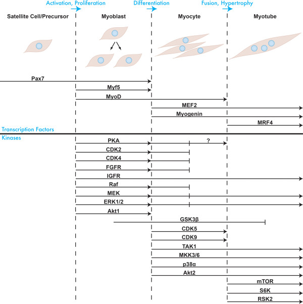
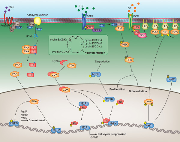
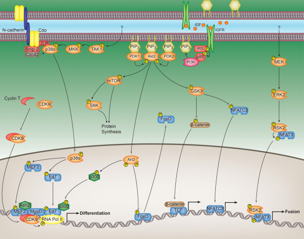
Myogenesis is a complex and tightly regulated process, the end result of which is the formation of a multinucleated myofibre with contractile capability. Typically, this process is described as being regulated by a coordinated transcriptional hierarchy. However, like any cellular process, myogenesis is also controlled by members of the protein kinase family, which transmit and execute signals initiated by promyogenic stimuli. In this review, we describe the various kinases involved in mammalian skeletal myogenesis: which step of myogenesis a particular kinase regulates, how it is activated (if known) and what its downstream effects are. We present a scheme of protein kinase activity, similar to that which exists for the myogenic transcription factors, to better clarify the complex signalling that underlies muscle development.


Figures

Figure 1
Figure 1
Transcription factors and kinases regulating the different stages of myogenesis. A graphical representation of myogenesis is shown. Embryonic precursors or quiescent satellite cells become activated to form proliferating myoblasts, which differentiate into myocytes that fuse to form a multinucleated myotube. The upper portion of the figure shows the myogenic transcription factors required for this process and the stages for which they are required. The lower portion shows the myogenic protein kinases and the stages that they regulate.
Figure 2
Figure 2
Regulation of the early myogenic transcriptional program by the kinome. The figure shows the mechanisms by which the kinases described in the text coordinate embryonic precursor activation, myoblast proliferation and the prevention of premature myoblast differentiation. Wnt1 and Wnt7a stimulation of precursor cells activates protein kinase A (PKA), which, through the phosphorylation of CREB, induces the expression of the myogenic transcription factors Myf5, MyoD and Pax3, resulting in the myogenic commitment of embryonic precursors. PKA then prevents the premature differentiation of proliferating myoblasts by phosphorylating and inhibiting the transcriptional activity of MEF2D. The cyclin-dependent kinases (CDKs) regulate cell cycle transitions and are activated at the appropriate time by the availability of their respective cyclins, depicted in the boxed inset. Cell cycle progression is achieved by the CDKs through the phosphorylation of Rb, which, when phosphorylated, is unable to bind and inhibit the E2F family of transcription factors that promote the expression of genes involved in cell division. Phosphorylation of Rb by the CDKs also prevents it from associating with and transactivating MyoD, thereby inhibiting cell cycle exit and differentiation. Cell cycle exit is further prevented by the proteolytic degradation of MyoD that results from direct CDK phosphorylation. The extracellular signal-regulated kinase (ERK) is activated by growth factors such as fibroblast growth factor and insulin-like growth factor (IGF), although the substrates ERK acts on to promote proliferation and inhibit differentiation are unknown. IGF also activates the Akt1 pathway and stimulates proliferation when myoblasts are subconfluent. Phosphorylation of FoxO1 by Akt1 prevents this transcription factor from accumulating in the nucleus, inhibiting the expression of genes involved in cell cycle exit, such as p27.
Figure 3
Figure 3
Regulation of the late myogenic transcriptional program by the kinome. The figure shows the mechanisms by which the kinases described in the text coordinate myoblast cell cycle exit, myoblast differentiation, myocyte fusion and myotube hypertrophy. Cell-cell contact and N-cadherin ligation, in conjunction with transforming growth factor-β-activated kinase 1 (TAK1) and MAP kinase kinase 3/6 (MKK), activate p38α. p38α induces cell cycle exit and differentiation through the phosphorylation of MEF2 and E47 that together with MyoD form part of an active myogenic transcriptional complex. A subunit of this complex is RNA polymerase II (RNA Pol II), which is phosphorylated and activated by cyclin-dependent kinase 9 (CDK9). Akt2, in response to IGF stimulation, phosphorylates the transcriptional coactivator and histone acetyltransferase p300, which is part of the same myogenic transcriptional complex. Activation of the Akt2 pathway promotes differentiation and hypertrophy by several other mechanisms as well. Akt2 phosphorylates and inactivates the FoxO family of transcription factors, whose activities are inhibitory to differentiation and hypertrophy. Phosphorylation of the mammalian target of rapamycin (mTOR) by Akt2 encourages protein synthesis/hypertrophy, partly through mTOR's phosphorylation and activation of the ribosomal protein S6 kinase 1 (S6K). Akt2 can also phosphorylate and inhibit glycogen synthase kinase 3β (GSK3). When active, GSK3 inhibits differentiation and hypertrophy through phosphorylation and cytoplasmic sequestration of NFATC3. Phosphorylation of β-catenin by GSK3 similarly prevents its nuclear accumulation and ability to activate the TCF/LEF family of transcription factors. Finally, activation of extracellular signal-regulated kinase 2 (ERK2) by an unknown stimulus promotes cell fusion through the phosphorylation and nuclear accumulation of NFAT3 via the 90-kDa ribosomal S6 kinase 2 (RSK2).

References

    1. Sabourin LA, Rudnicki MA. The molecular regulation of myogenesis. Clin Genet. 2000;57:16–25. - PubMed
    1. Le Grand F, Rudnicki MA. Skeletal muscle satellite cells and adult myogenesis. Curr Opin Cell Biol. 2007;19:628–633. doi: 10.1016/j.ceb.2007.09.012. - DOI - PMC - PubMed
    1. Manning G, Whyte DB, Martinez R, Hunter T, Sudarsanam S. The protein kinase complement of the human genome. Science. 2002;298:1912–1934. doi: 10.1126/science.1075762. - DOI - PubMed
    1. Caenepeel S, Charydczak G, Sudarsanam S, Hunter T, Manning G. The mouse kinome: discovery and comparative genomics of all mouse protein kinases. Proc Natl Acad Sci USA. 2004;101:11707–11712. doi: 10.1073/pnas.0306880101. - DOI - PMC - PubMed
    1. Walsh DA, Perkins JP, Krebs EG. An adenosine 3',5'-monophosphate-dependant protein kinase from rabbit skeletal muscle. J Biol Chem. 1968;243:3763–3765. - PubMed

LinkOut - more resources